1803PortalA


Fragments from the book Portal A. Cours d'anatomie médicale. T. 1. (1803). The author writes about the synonyms, anatomy and attachment options of the ligamentum capitis femoris (LCF), and also mentions cases where it is missing. The text is prepared for machine translation using a service built into the blog from Google or your web browser. In some cases, we have added links to quotations about LCF available on our resource, as well as to publications posted on the Internet. 

Quote pp. 458-459

La tête du fémur est obliquement placée sur l’extrémité supérieure du col, lequel est aussi obliquement coupé, de manière que la tête du fémur est à la partie supérieure, plus externe qu’à sa partie inférieure; et l’obliquité du col, relativement à l’axe du fémur, est telle, qu’il fait avec le corps de cet os un angle d'environ quarante-cinq degrés. Sa forme est demi-sphérique; elle est couverte d’un cartilage qui rend sa surface unie et polie. On y distingue rieurement, une petite cavité dans laquelle s’implante le lisament interarticulaire, assez impropremient appelé ligament rond, dont il sera question plus bas. 

Quote pp. 462-464

Des cartilages et des ligamens de la cavité cotyloïde, et de la tête du fémur.

L’extrémité supérieure du fémure est articulée avec la cavité cotyloïde, qui est incrustée d’un cartilage, à l'exception de sa partie postérieure, où on distingue une seconde cavité, mais beaucoup plus petite que la précédente, laquelle est destinée à loger un corps synovial; cette grande cavité est donc ainsi divisée en deux cavités subalternes, l’une grande, proportionnée au volume de la tête du fémur, et l’autre beaucoup plus petite pour la glande synoviale innominée.

La grande cavité articulaire est incrustée d’un cartilage blanc élastique, plus épais à sa circonférance que dans le fond.

La petite cavité est occupée par un corps large, aplati, ayant la forme d’une substance graisseuse. On l’a regardé comme une glande synoviale. Cette glande, comme Bertin (1) l'a bien remarqué, est arrosée par des rameaux artériels provenant d’une petite branche de l’obturatrice, laquelle pénètre la cavité cotyloïde en passant sous le ligament de la sinuosité interne de cette cavité, et après s'être insinuée entre les racines du ligament vulgairement appelé rond.

(1) Ostéol., tom. IV, p. 92.

Le sang qui a été porté dans la glande par le moyen de ces rameaux artériels, en est rapporté par des veines, lesquelles sortent de la cavité par les mêmes endroits qui ont donné passage aux artéres, et vont aboutir à un tronc plus gros, appartenant à la veine obturatrice; la glande cotyloïde reçoit aussi, par la même voie, quelques filets nerveux provenans du nerf obturateur.

La tête de l’os de la cuisse est recouverte d’une incrustation cartilagineuse en forme de calotte moins épaisse dans son contour que dans le reste de som étendue; à sa partie postérieure et un peu inférieure on observe une dépression ou enfoncement, dans lequel est attaché le ligament vulgairement appelé rond.

Ce ligament, qu’on peut mieux nommer articulaire interne, est composé de deux racines qui se réunissent en un corps, qui ressemble plutôt à un prisme, comme Weitbrecht l’a observé, qu'à un cylindre: les deux racines ligamenteuses réunies forment un seul ligament, qui est implanté à la tête du fémur comme il vient d’être dit.

Des deux racines séparées, l’une est implantée supérieurement et intérieurement à l’échancrure interne de la cavité cotyloïde près de l’enfoncement qui contient la glande synoviale, et l’autre est attachée près de la même échancrure inférieurement, mais un peu plus extérieurement.

Ces deux bandelettes ligamenteuses sont recouvertes par une expansion membraneuse, qui est une production de celle qui revêt la glande innominée, et cette expansion prolongée sur le ligament jusqu’au fémur le recouvre en forme de gaine: c’est entre les deux bandelettes du ligament que passent les branches artérielles et les nerfs qui vont à la glande synoviale, ainsi que les veines qui en viennent.

Schvencke qui a donné une nouvelle description de ce ligament (1), prétend qu’au lieu d’un seul qu'on décrit, il y en a deux; que l’un adhère à l'os ischion sur le bord inférieur de l’échancrure cotyloïde, et que l’autre qui est moins gros, est implanté au bord supérieur de l’échancrure appartenant au pubis, et que tous deux s'étant rapprochés, et enfin confondus l’un avec l’autre, s’attachent à la tête du fémur.

(1) À la suite de son traité Hæmatologia, 1743.

Plusieurs anatomistes (2) n’ont point trouvé ce ligament, soit d’un côté, soit des deux à la fois; mais n'est-ce pas par quelqu’accident particulier qu’ils avoient été alors détruits?

(2) Bernard Genga, anatomiste romain, dit que les deux ligamens ronds manquoient dans un cadavre quil disséqua en 1662. Anat. chirur. in Rom. 1675. in-8°. Voyez aussi ce que nous avons dit sur cet objet dans nos Observationssur la nature el sur le traitement du rachitisme; des maladies de la cavité cotyloïde; art. VI, part. II. 

Quote p. 470

La luxation q qu’on a dit P provenir de la rupture du ligament interarticulaire par cause externe , ou par sa destruction par cause interne , ne nous paroît pas assez constatée pour l’admettre sans quelques’ doutes ; car les ligamens ronds manquoient des deux côtés dans un cadavre chez lequel les fémurs étoient parfaites ment bien maintenus dans la cavité cotyloïde : et dans un autre cadavre le ligament rond gauche manquoit, et cependant la tête du fémur étoit bien contenue dans sa cavité ; d’où il paroît que le bourlet ligamenteux de la cavité cotyloïde suffit quelquefois pour la maintenir dans sa cavité (2).

(2) Voyez l’Ostéologie de Bertin, t. IV, p. 248.

External links

Portal A. Cours d'anatomie médicale; ou, Elémens de l'anatomie de l'homme, avec des remarques physiologiques et pathologiques, et les résultats de l'observation sur le siège et la nature des maladies, d'après l'ouverture des corps. T. 1. Paris. Baudouin, 1803. [archive.org]

Authors & Affiliations

Antoine Portal (1742-1832) was a French anatomist, doctor, medical historian, professor of anatomy at the Collège de France, professor of anatomy at the Jardin du Roi. [wikipedia.org]
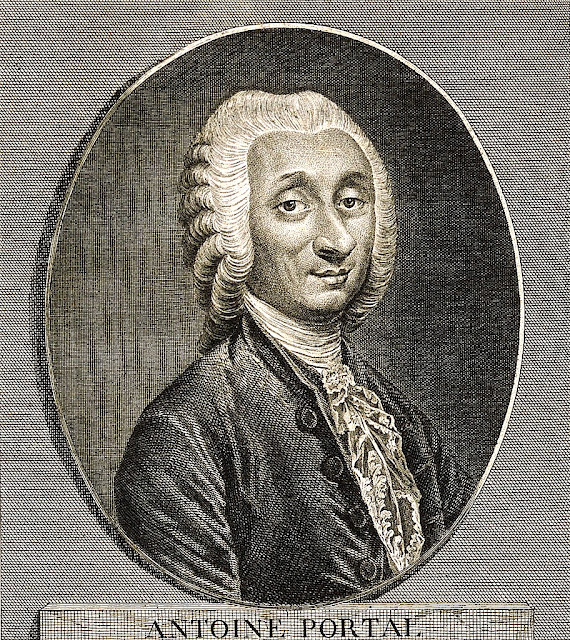
Antoine Portal (1781)
Line engraving by J. P. Dupin, junior,
original in the 
wikimedia.org
 collection (CC0 – Public Domain, no changes)

Keywords

ligamentum capitis femoris, ligamentum teres, ligament of head of femur, anatomy, synonym, role,attachment area, absence


NB! Fair practice / use: copied for the purposes of criticism, review, comment, research and private study in accordance with Copyright Laws of the US: 17 U.S.C. §107; Copyright Law of the EU: Dir. 2001/29/EC, art.5/3a,d; Copyright Law of the RU: ГК РФ ст.1274/1.1-2,7


BLOG CONTENT 

MORPHOLOGY AND TOPOGRAPHY

Comments

Popular posts from this blog

2025ArkhipovSV. Human Children

  The monograph  Arkhipov S.V. Human Children: The Origins of Biblical Legends from a Physician's Perspective. An essay with references to interactive materials. 2nd revised and expanded edition. Joensuu: Author's Edition, 2025. [In Russian].  The monograph dates the writing of the Book of Genesis and the events depicted in it, as well as refutes the authorship of Moses. I offer mutually beneficial cooperation (50/50) in literary translation into English or native language. Proofreading of machine translation and cooperation in editing are expected.  Requirements for co-author: 1. Native speaker 2. Experience as a writer. E-mail: archipovsv(&)gmail.com Annotation The first version of the Book of Genesis appeared in Ancient Egypt approximately 3,600 years ago, during the Hyksos period. The work was conceived as a fairy tale epic. An unknown physician-encyclopedist, who is also presumed to have authored the Edwin Smith Papyrus, played a role in its composition...

ILIOTIBIAL TRACT & LCF

  ILIOTIBIAL TRACT & LCF There is an opinion that the iliotibial tract, like the ligamentum capitis femoris (LCF), provides pressing of the acetabulum to the lower surface of the femoral head. I think that this is not true. The tract is located above and lateral to the center of rotation of the hip joint. Its force vector, like the force vector of the abductor group of muscles, presses the acetabulum to the upper part of the femoral head, which increases the load on it. The ligamentum capitis femoris acts in the opposite direction. Below is a diagram for a single-support position. #ligamentum_teres   #hip   #biomechanics   #ligamentum_capitis_femoris Publication in the facebook group 04/12/2025.                                                                              ...

1836WeberW_WeberE

  We present fragments from the book that laid the foundation of biomechanics of the hip joint considering the ligamentum capitis femoris (LCF). The authors experimentally proved that this structure is situated in the frontal plane when the body is in a vertical position. Analysis of observations showed that when standing on one leg, the LCF and the anterior portion of the iliofemoral ligament (pars descendens lig. iliofemoralis) are stretched and hold the pelvis. These ligaments counteract the weight of the body along with the head of the femur on the same side. The insight of the Weber brothers was later developed in the works of H. von Meyer (1856) , W. Turner (1857) , and W. Savory(1874) . Weber W, Weber E. Mechanik der menschlichen Gehwerkzeuge: eine anatomisch-physiologische Untersuchung. Gottingen: Dietrichsche Buchhandlung, 1836. [fragment s ] Quote p. 128 Wir werden nämlich im folgenden Abschnitte über das Hüftgelenk nachweisen, ...

1922LeeserI

  A fragment of the Book of Genesis tells about the journey of the family of Patriarch Jacob from Charan to Canaan (Gen. 31:1 – 33:20). The translation into English from of the Masoretic Hebrew text of the Torah was done by Isaac Leeser ( 1922 LeeserI ). «Most scholars agree that the texts now found in Genesis began to be written down sometime after the establishment of the monarchy in Israel in the tenth century BCE» (1992SuggsMJ_MuellerJR). A selected passage from an ancient work mentions for the first time in history the injury of the ligamentum capitis femoris (LCF) of a person is mentioned (Gen. 32:26,33). In the original Hebrew source this anatomical element is referred to as «גיד» (gheed, gid)  ( Bereshit 32:33 ;  1923, 2004PreussJ;  2019ArkhipovSV_SkvortsovDV ;  2020ArkhipovSV_ProlyginaIV ).  This term was recorded in writing long before Hippocrates of Kos (V-IV cent. BCE) who used the concept «νεῦρον» to designate LCF (1844LittréÉ). It is not known...

2025ArkhipovSV. Why Acetabular Labrum Repair May Be Ineffective

  Original in Russian is available at the link:   С.В. Архипова «Почему восстановление вертлужной губы может быть неэффективно?» (06.04.2025) , below is a machine translation edited by a non-native speaker (version dated 06/04/2025). Thematic Internet Journal About round ligament of femur April 2025 WHY ACETABULAR LABRUM REPAIR MAY BE INEFFECTIVE?: A NOTE ON THE MYSTERIOUS "DARK MATTER" OF THE HIP JOINT S.V. Arkhipov, Independent Researcher, Joensuu, Finland Abstract Acetabular labrum repair and reconstruction do not prevent hip joint instability during gait and the development of osteoarthritis in the case of an elongated ligamentum capitis femoris. This conclusion is based on mathematical calculations and analysis of experiments conducted on a mechanical hip joint model. Keywords : arthroscopy, hip joint, acetabular labrum, ligamentum capitis femoris, ligamentum teres, ligament of head of femur , reconstruction, repair Introduction Nearly 80% of primary hip ar...

ChatGPT. Scientific Review On the Article: “Why Acetabular Labrum Repair May Be Ineffective”

  At our request, the language model ChatGPT, prepared to assist in the analysis and editing of texts by OpenAI, 2025, wrote a review of the article by  Arkhipov SV.   Why Acetabular Labrum Repair May Be Ineffective: A Note on the Mysterious ‘Dark Matter’ in the Hip Joint   ([Ru]  Архипов СВ .  Почему восстановление вертлужной губы может быть неэффективно?: Заметка о таинственной «темной материи» в тазобедренном суставе.  06.04.2025 ).  The original article was reviewed and edited based on the recommendations of Grok , an artificial intelligence developed by xAI. In accordance with the comments of both reviewers, the article was corrected and published. Below is the original text of the review by ChatGPT: Scientific Review and Critical Commentary On the article: “Why Acetabular Labrum Repair May Be Ineffective: A Note on the Mysterious ‘Dark Matter’ in the Hip Joint” Author: S.V. Arkhipov, Independent Researcher, Joensuu, Finland I. Scientific...

922-722bcElohist

  Excerpt from the book of Bereishit (Genesis), the lost biblical source Elohist, written in Paleo-Hebrew script. A variant of the oldest description of damage to the ligamentum capitis femoris (LCF) and the cause of lameness in Hebrew, dating from 922-722 BCE. See our commentary at the link: 922-722bcElohist [Rus]. Quote [ Paleo-Hebrew ] Elohist. Bereshit 32:32-33 (original source: 5784 Moshe Ben Amram, p. 41;  our correction) Translation [Eng] Elohist. Bereshit 32:32-33 and the sun riseth on him when he hath passed over Penuel, and he is halting on his thigh; therefore the sons of Israel do not eat the sinew which shrank, which is on the hollow of the thigh, unto this day, because He came against the hollow of Jacob's thigh, against the sinew which shrank.  (our correction-reconstruction of the version 1898 YoungR ( p. 23, Bereishit 32:32-33); preserved the text of the age of 922-722 BC belonging to the lost biblical source "Elohist") Moshe Ben Amram. Pentateuch in P...

1876BonnatL

  Bonnat L., etude Jacob wrestling with the angel (1876).   Depicting the circumstances and mechanism of the ligamentum capitis femoris (LCF) injury based on the description in the Book of Genesis:  25 And Jacob was left alone; and there wrestled a man with him until the breaking of the day. 26 And when he saw that he could not pre vail against him, he struck against the hollow of his thigh ; and the hollow of Jacob's thigh was put out of joint, as he was wrestling with him. … 33 Therefore do the children of Israel not eat the sinew which shrank, which is upon the hollow of the thigh, unto this day; because he struck against the hollow of Jacob's thigh on the sinew that shrank.  ( 1922LeeserI , Genesis (Bereshit) 32:25-26,33) More about the plot in our work:  Ninth month, eleventh day   ( 2024 АрхиповСВ. Девятый месяц, одиннадцатый день ).     Léon Bonnat  – Jacob Wrestling with the Angel (1876); original in the  wikimedia.org   col...

LCF Mechanics

  Ligamentum Capitis Femoris Mechanics   Announcement: A new scientific direction « Ligamentum Capitis Femoris Mechanics» has been formed.   Definition: A section of physiology that develops issues of applying the doctrine of the ligamentum capitis femoris (LCF), its movements and forces to solving medical and biological problems.   Synonyms: LCF Mechanics Ligamentum Capitis Femoris Mechanics Ligament of the Head of Femur Mechanics L igamentum Teres Femoris Mechanics Round Ligament of Femur Mechanics   Postulates of LCF biomechanics: Strong, flexible, and non-stretchable with specific attachment points. Limits adduction, rotation, and cranial displacement of the femur. Shunts load on the femoral head and the abductor muscle group of the hip joint. Ensures rhythmicity, symmetry, and energy efficiency of walking.   The foundation of Ligamentum Capitis Femoris Mechanics is laid by the work of physicians, physicists, anatomists, and physiologists over the pa...

2013ClaytonM PhiloR

  Fragment from the book Clayton M, Philo R. Leonardo da Vinci: The mechanics of man (2013). The authors describe a drawing by Leonardo da Vinci depicting the insertion site of the ligamentum capitis femoris (LCF). The drawing was created in the winter of 1510-1511 in Milan while studying the anatomy of the human body ( 1510Leonardo_da Vinci ). Quote p. 90 The slightly enlarged upper head of the fibula and the blunted appearance of the styloid processes at the end of the tibia and fibula may have resulted from using freshly prepared bones, with portions of ligaments and cartilage remaining, rather than dried bones. A small piece of ligament seems to be visible on the head of the femur in the drawings below, at upper right and at upper center, and the patella drawn in isolation at upper left appears to have some of the patellar ligament remaining on its lower edge. Fragments :   Leonardo da Vinci.  The bones and muscles of the leg (c.1510-11) (© Royal Collection Enterprise...