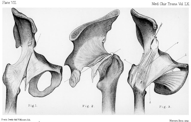
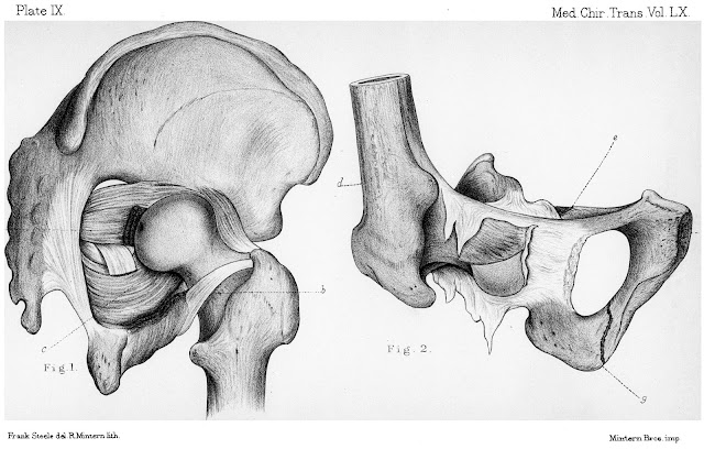
An excerpt from the article by Morris H. Dislocations of the Thigh: their mode of occurrence as indicated by experiments, and the Anatomy of the Hip-joint (1877, pp. 161-170). The author noted that the ligamentum capitis femoris (LCF) is stretched during flexion, adduction, and external rotation. It was found that the LCF is always damaged in hip dislocations, is more often torn from the femur, and is rarely ruptured. Discussion of article see article 1877BrookeC.
DISLOCATIONS OF THE THIGH: THEIR MODE OF OCCURRENCE AS INDICATED BY
EXPERIMENTS AND THE ANATOMY OF THE HIP-JOINT.
BY HENRY MORRIS, M.A., M.B., F.R.C.S., ASSISTANT SURGEON TO, AND
LECTURER ON ANATOY1 AT, THE MIDDLESEX HOSPITAL.
Received January 9th-Read February 13th, 1877.
RECENTLY, whilst making various dissections of the hip-joint, the
usually accepted doctrine that dorsal and ischiatic dislocations of the femur
occur when the thigh is in a position of adduction appeared to me as very improbable,
on anatomical grounds alone, and I was, therefore, led to test the truth of it
my means of experiments on the dead body.
I propose in the following remarks to prove by anatomy, experimental
results, and clinical facts-(I) that all kinds of dislocations at the hip-joint
can take place while the thigh is abducted; (2) to give reasons for believing
that abduction is the position in which all dislocations of the thigh happen;
and (3) to show that in any given case the dislocation will be backwards, downwards,
or forwards, according as flexion with rotation inwards, or extension, or
extension with rotation outwards, is associated with abduction at the moment of
accident; or is provoked by the same violence which produces the displacement.
First, I will state briefly the anatomical features which bear upon the
question.
The acetabulum looks forwards as well as outwards and downwards, and
receives the head of the femur, which also looks forwards. The thickest and
strongest part of the innominate bone is that which forms the upper and posterior
wall of the acetabulum; it is this part of the bone which enters into the
formation of the weight-bearing arches of the pelvis, viz. of the pelvic brim, along
which the weight is transmitted to the heads of the thigh bones in the upright
and stooping positions, and of the vertical or ischial arch, along which the
weight is transmitted to the tuberosity of the ischium in the sitting posture.
This same part of the innominate bone forms by far the deepest portion of the
acetabulum, the articular surface of which is fully an inch wide in its iliac
portion, and nearly as wide in the ischial. Along the lower part of the
acetabulum, on the other hand, the articular facet, so far as it exists at all,
is very narrow; while the cotyloid notch intercepts the rim for nearly one inch.
The head of the femur presents a much larger articular surface above the
transverse plane through its centre than below it. The measurement (in a
horizontal plane), from a vertical line skirting the dimple for the round ligament
to the outer limit of the articular surface on the upper aspect of an average
femur, is an inch and two thirds; whereas to the outer border of the articular
surface below is only three eighths of an inch. The notch for the round ligament
is in the lower and posterior quarter of the head; the most prominent point in
the head is below this notch, while the part above slopes off from within outwards.
As a consequence of all this, when the thigh is flexed and adducted, the most
projecting part of the head passes into the deepest part of the acetabulum and presses
against the broadest and strongest part of its wall; whereas in abduction this
prominent part of the head of the femur projects beyond the lower part of the socket
at the cotyloid notch.
THE LIGAM1ENTS. It is only necessary to refer to two of the four
ligaments of the joint, viz. the capsular and the round ligament.
The capsular ligament is large and loose, so that in every position of
the limb some portion of it is relaxed. In thickness and strength it varies
greatly in different parts; thus if two lines be drawn, one from the anterior inferior
iliac spine to the inner border of the femur near the small trochanter (Plate
VIII, fig. 1), the other from the upper border of the tuber ischii to the
digital fossa (Plate VIII, fig. 2), the ligament above and behind these lines,
is very strong; whereas all below, except along the narrow pectineo-femoral
band, is very thin and weak, sometimes permitting the head of the bone to be
seen through it. There are three bands auxiliary to the capsule, or more correctly
speaking, three portions of the capsule, which have received separate names,
and which for the sake of description are called ligaments, although it would
be erroneous to suppose that either of these bands or ligaments are structures
separate from the capsule. The best known of them is the ilio-femoral ligament,
a large triangular mass of coarse, well-marked fibres, the broadest end of
which is at the femur (Plate VIII, fig. 3). Its fibres are somewhat accumulated
both along the outer and inner sides of the femoral attachment, while several
curved intercolumnar fibres, which spring from the centre of the capsule, pass
to the femur with the inner set.
This arrangement of the fibres has led to the ligament being called by
some the "inverted Y-shaped ligament," and exaggerated
representations of it, in accordance with this name, have been drawn. Such, for
instance, are the diagrams of the ligament in Bigelow's work and in the works
of those who have copied therefrom. The name is, however, inappropriate, for
although the double accimulation of fibres may be made, by dissection, to show
somewhat the appearance of an inverted Y, there is not any natural interval
between the two imaginary arms of the ligament beyond a small aperture for the
entrance of a branch of the external circumflex artery into the joint. Except
for this small opening, the strong central fibres which take a straight course
to the femur are not interrupted, and are of considerable thickness. The
mistake has arisen, I imagine, from the presence of two well-marked bands
connected with the surface of the capsule along the upper and outer border of
the ilio-femoral ligament. One of these, the shorter and posterior, passes to
the tendon of the gluteus minimus from the middle of the capsule; the other,
not hitherto described, but generally well marked, extends from the vastus
externus to the long head of the rectus femoris, and moves with both these
tendons (Plate VIII, fig. 3). The latter especially gives the appearance of a superadded
outer arm to the ilio-femoral ligament, while the more oblique direction of the
inner fibres completes the supposed Y-shaped outline.
The ischio-femoral ligament extends from the ischial border of the
acetabulum to the upper and back part of the neck of the femur, just internal
to the highest part of the digital fossa. When the thigh is flexed the fibres
of this ligament pass almost straight to their femoral insertion, and are
spread out uniformly over the head of the femur, but in extension they wind
upwards and outwards to the femur in a zonular manner (Plate VIII, fig. 2), and
form quite a thick roll along their lower border. The upper and back part of
the capsule between the ilio-femoral and ischio-femoral ligaments is very
strong and thick, although not so strong and thick as the ilio-femoral itself.
The latter is as stout as the tendo Achillis, and is often more than a quarter
of an inch in thickness; the rest of the strong part of the capsule varies down
to one eighth of an inch or a little less in thickness.
The third auxiliary band is the pectineo-femoral ligament (Plate VIII,
fig. 3), a distinct but narrow set of fibres, extending from the pubis and
pubic border of the cotyloid notch to the lower and back part of the neck of
the femur just above the small trochanter.
The effect of these bands on movements of the hip-joint is as follows:
The ilio-femoral supports the trunk in the erect posture by limiting extension;
it is made tense throughout in every position of extension, except when extension
is combined with abduction, and then the outer fibres are relaxed.
The ischio-femoral band does not limit simple flexion, but is tight
during flexion combined with adduction, and flexion combined with rotation
inwards. In extension, in flexion with rotation outwards, and in abduction, it
is quite loose. The part of the capsule between the ilio- and ischio-femoral
ligaments assists the former in limiting simple extension; it is also made
tense in extension with adduction, but is most tense in adduction combined with
slight flexion, - the stand-at-ease position.
The pectineo-femoral ligament is stretched in every position of flexion
and of extension when associated with abduction. It is the structure which
limits abduction, and is especially tight during abduction combined with slight
flexion, and also when marked flexion with outward rotation accompanies
abduction.
The ligamentum teres, so far as its attachments are concerned, needs no
notice here, but its action and use require a word or two. Without discussing
the differences of opinion which have been expressed on these points it will be
sufficient to state, as the result of several examinations, that the ligament
is most tight during flexion combined with adduction and rotation outwards, but
that it is also tight, though to a less degree, during flexion with rotation
outwards, as well as during flexion with adduction. As the thigh passes from a
position of adduction with flexion to one of adduction with extension it
becomes less and less tense, and is quite lax when full extension is reached.
In every position of abduction it is loose, while in abduction associated with
simple flexion it is at its loosest, and the ligament is then folded upon
itself so completely as to have its femoral and acetabular attachments on the same
level and opposite to one another.
From a consideration of these anatomical facts, it is evident that no
conditions of the joint can be more favorable to rupture of the capsule and
dislocation of the thigh than those of abduction. In abduction the head of the femur
is more than half out of the acetabulum, bulging over the transverse ligament;
the cotyloid notch being filled with soft compressible fat allows the ligament
to yield a little as the head of the bone is forced over it; and the most
prominent part of the head of the femur is resting hard against the thin
portion of the capsule. This part of the capsule is strengthened somewhat by
the pectineo-femoral ligament it is true, but it derives no appreciable support
from muscles as does the rest of the capsule. The ligamentum teres, being quite
loose, is absolutely powerless to resist force, but is most favorably situated
to snap under a sudden jerk. Finally, in abduction all the strong portion of
the capsule is relaxed, excepting during complete extension, when the innermost
fibres of the ilio-femoral ligament become tense. In fact, it may be 8aid that
there is a natural tendency for the head of the femur to be displaced during
abduction of the thigh.
Nor is there any other position so favorable to dislocation as
abduction. It is quite unnecessary to discuss the improbability of dislocation
in positions of simple extension, when the ilio-femoral ligament is the
resisting structure; it is only in the rarest, i.e. the so-called irregular
dislocations, that this part of the capsule is ever ruptured, and even then
only by the extension of the laceration commenced elsewhere, and the peeling
off of the ligament from its bony connections.
There is, however, a pretty prevalent opinion that dorsal dislocations take
place during adduction through a rent in the upper and back portion of the
capsule, and that the use of the round ligament is to resist forces which tend
to drive the bone out in this direction. Now, as dorsal dislocations are more
common than all the others put together, it is most likely that they occur
through some strain upon the weak part of the capsule, and it is necessary to
state the anatomical provisions against their occurring during adduction. It is
only during flexion with marked adduction, and flexion with rotation inwards, that
the head of the femur presses firmly against the upper and back part of the
capsule, and even in these positions the most prominent part of the head is
against the strongest and deepest side of the acetabulum. Daring flexion with
adduction the ligamentum teres is on stretch, and therefore in action to resist
displacing forces, so far as it ever can do so. The portion of the capsule
against which the head of the bone is bulging is strengthened by various
muscles, and these during adduction and inward rotation lie stretched over it;
the most important of them is the obturator internus, which, when stretched,
acts much like a ligament on account of the several tendons into which the
muscular tissue is inserted and the connection of some of the ultimate fibres
of these tendons with the bony origin of the muscle; the power of resistance of
this muscle is increased too by the play of the tendons of the trochlear groove
of the ischium, whereby the strain on the muscle is diminished. Finally, the
close connection between the external rotator muscles, as they pass to their
insertions, and also of the gluteus minimus, with the capsule, must strengthen
and support the upper and back part of that ligament.
Thus, the anatomy of the joint, it seems to me, points to abduction as
the only position in which dislocation of the thigh can possibly occur.
Experiments upon the dead body confirm the conclusion suggested by anatomy. They
also establish the fact that all the regular varieties of dislocation of the
hip can be produced and reduced after the head of the femur has been displaced
in abduction. It will be best at once to state that the only way in which I
have been able to dislocate the thigh is by forcibly abducting it. I have
tried, standing over the body, the pelvis of which has been firmly fixed by two
men, to force the bone out of the acetabulum through the back of the capsule; I
have removed all the muscles and tried the same thing when only the capsule and
wall of acetabulum were left to resist, and when, by previously flexing and
adducting, or flexing and rotating inwards the thigh, the strain upon the
capsule is at its greatest. I have also tried, by bringing the legs across one
another, and jerking and forcing the thigh with the whole leverage of the limb
to assist me, as well as by striking with a heavy block the front and outer
side of the knee, to dislocate the flexed and adducted thigh; but by none of these
means have I ever once succeeded.
As soon, however, as the limb is abducted and jerking force is applied
to it while in that position, the head of the femur bursts through the capsule,
and the dimple for the round ligament can be felt beneath the skin of the perinaeum.
After the head has left the acetabulum it can be made to pass either towards
the dorsum ilii or the ischiatic notch by flexing and rotating the limb inwards
more or less; or on to the pubis by extending and rotating it outwards.
The following statements are based on fifteen experiments:
The results in all these cases were so similar that it seemed
unnecessary to record the notes of more. Except when specially stated to the
contrary all the dislocations were effected before any dissection whatever was
commenced.
1. The rent in the capsule varied with the degree to which the upward
displacements had been carried previous to dissection, but it was always
limited to the thin portion of the capsule. In none did it involve the
ilio-femoral or the ischio-femoral ligament, or the strong part of the capsule
between them. In most cases it commenced at the pubis, ran obliquely downwards
and backwards to the neck of the femur, and then extended upwards to a greater
or less distance towards the digital fossa, either peeling the capsule from the
back of the neck of the femur or tearing it through at a short way from its attachment
to that bone (Plate VIII, figs. 1, 2). In some cases the periosteum was torn
away with the ligament from the pubic part of the acetabulum, and in one the
whole of the under and inner side of the capsule was separated from the
acetabular margin; but in all, the rent took the same general course along the
femoral insertion of the capsule.
2. The ligamentum teres was ruptured in every case by the dislocating
force, and not by the subsequent movements whereby the dorsal or other
positions were obtained. This was ascertained by feeling for the dimple in the
head through the periraeum. In all but three the ligament was detached from the
femur, and with it, in some instances, a scale of articular cartilage. In three
cases it was torn asunder, once near the femur and twice near its middle.
3. The pectineus, quadratus femoris, and external obturator muscles were
ruptured, the two former in every case, the latter each time it was examined.
The gemellus inferior was generally torn asunder.
4. The obturator internus was ruptured in two instances, and the
adductors longus and brevis in one and the same case. In none was the neck of
the femur embraced by the small rotator muscles (viz. gemelli, obturator
internus, and pyriformis); nor could the head of the bone be made to pass
upwards beneath the obturator internus tendon, i. e. between it and the capsule
of the joint, but it could be forced upwards onto the dorsum over the posterior
surface of the muscle.
5. In no case was the ilio-psoas or the gluteus minimus ruptured. In one
of the cases in which the obturator internus was torn the pyriformis and the
lower border of the gluteus medius were also lacerated.
6. In three cases the great sciatic nerve was lying in front of the neck
of the bone, and was not seen in the dissection of the gluteal region until the
dislocation was reduced. In one case the nerve was carried before the neck and
retained there after reduction.
7. In three cases the dislocation' was compound,' so that the head of
the femur protruded through the skin of the perinaeum.
…
External links
Morris H. Dislocations of the Thigh: their mode of occurrence as indicated by experiments, and the Anatomy of the Hip-joint. Medico-Chirurgical Transactions. 1877;60:161-186.1. [pmc.ncbi.nlm.nih.gov]
Brooke C. Dislocations of the Thigh: their mode of occurrence as indicated by experiments, and the Anatomy of the Hip-joint. By Henry Morris. M.A., M.B. Reports of Societies. Br Med J. 1877Feb17;1(842):203. [pmc.ncbi.nlm.nih.gov]
Authors & Affiliations
Henry Morris (1844-1926) was a British medical doctor and surgeon. [wikipedia.org]
 |
| Sir Henry Morris (before 1915) Author Anton Mansch, published by A. Eckstein, Berlin; original in the wikimedia.org collection (CC0 – Public Domain, no changes). |
Keywords
ligamentum capitis femoris, ligamentum teres, ligament of head of femur, role, dislocation, injury, damage
NB! Fair practice / use: copied for the purposes of criticism, review, comment, research and private study in accordance with Copyright Laws of the US: 17 U.S.C. §107; Copyright Law of the EU: Dir. 2001/29/EC, art.5/3a,d; Copyright Law of the RU: ГК РФ ст.1274/1.1-2,7


Comments
Post a Comment