
Primates: Prosimii & Anthropoidea
In the Late Devonian, or 367.5 Ma, the Amniota branch arose from amphibians (2011PyronRA). In that cohort, synapsids (Synapsid) initially emerged, followed by sauropsida (Sauropsida), and then reptiles (Reptilia) (1995LaurinM_ReiszRR). The first representatives of the reptile-like (Reptilomorpha) tetrapods colonized the continents 363-290 Ma (1995LundbergJG).
The earliest synapsid is recognized as Asaphestera platyris from the early Pennsylvanian subperiod, which lasted from 323.2 ± 0.4 to 315.2 ± 0.2 Ma (2020MannA_AndersonJS; 2023CohenKM_CarN). According to molecular clock data, mammals (Mammalia), also called animals (Theria), separated 310 Ma from the lineage that extended to reptiles (2004HedgesSB_ShoeJL). The first animals that suckled their young with milk stood out among the primitive synapsids - the ancestors of cynodonts (Cynodontia) (2013VaughanTA_CzaplewskiNJ). The oldest mammal Liaoconodon hui was found in deposits of the early Cretaceous period, that is, it lived 145.0-100.5 Ma (2011MengJ_LiC; 2023CohenKM_CarN).
The
molecular clock «shows»: the superorder Euarchontoglires separated in the class
of mammals 88.8 Ma (2007JaneckaJE_MurphyWJ). This systematic unit, also called
superprimates (Supraprimates), is subdivided into: the grandorder Glires with
the orders Lagomorpha and Rodents; the grandorder Euarchonta with the orders
Scadentia and the worldorder Primates (Primatomorpha), in which the orders
Dermoptera and Primates («Leaders of Creation») are distinguished
(2017EsselstynJA_FairclothBC).
 |
| Light-fronted spider monkey; exhibit of the Zoological Museum of Moscow State University, photo by the author. |
Molecular evidence suggests that euarchonts evolved 87.9 Ma, primates 86.2 Ma, great apes 79.6 Ma, and tree shrews 63.4 Ma (2007JaneckaJE_MurphyWJ). According to the fossil record, placental mammals diversified about 66 Ma, and the earliest plesiadapiform primate lived 65 Ma (2015ChesterSG_ClemensWA). The oldest remains of a definite primate, Teilhardina asiatica, were found in soil strata of the early Eocene, or 55.5 million years old (2006SmithT_GingerichPD; 2023CohenKM_CarN).
Apes emerged in the animal kingdom approximately 50 Ma (2009SrivastavaRP). One of the original hominoids, Morotopithecus bishop, flourished in Africa 20.6 Ma (1997GeboDL_PilbeamD). Its close «relative» primate Afropithecus lived 17.5-17.0 Ma (1997LeakeyM_WalkerA). According to molecular estimates, orangutans differentiated 19.3-15.7 Ma, gorillas - 9.7-7.6 Ma, and chimpanzees - 6.5-5.8 Ma (2011IsrafilH_SteiperME). Evolutionists believe that gibbons separated 29.62-20.68 Ma, orangutans - 18.42-12.53 Ma, gorillas - 9.89-6.62 Ma, common chimpanzees - 6.52-4.77 Ma, pygmy chimpanzees (bonobos) - 5.85-4.35 Ma (2022PoszewieckaB_GambinA).
Currently, the order of primates includes 190
species, placed in the suborder lower primates (Prosimii) with the families:
tupaiids (Tupaiidae), lorises (Lorisidae), lemurs (Lemuridae); and in the
suborder of higher primates (Anthropoidea) with the families: prehensile-tailed
monkeys or capuchins (Cebidae), marmosets (Cercopithectidae), great apes
(Pongidae) and hominids (Hominidae) with the only species, Homo sapiens
(1979NaumovNP_KartashevNN). In Africa and Asia, seven species of great apes of
three genera still survive: orangutan (Pongo), gorilla (Gorilla) and chimpanzee
(Pan) (2005WilsonDE_ReederDM). Today, orangutans and gorillas make up the
subfamily hominins (Homininae), and humans and chimpanzees are united in the
tribe Hominini, as having descended from a common ancestor (2001GrovesCP).
The presence of ligamentum capitis femoris
(LCF) in an animal can be determined based on the analysis of the acetabulum
and proximal femur. On the femur, these are the fossa of the femoral head, the
groove of the femoral head, the tuberosity or cleft on the femoral head, and the
marginal defect of the articular surface of the femoral head. In the pelvic
area, the presence of LCF is indicated by: the acetabular notch, the acetabular
fossa, the opening of the acetabular floor, and irregularities on the articular
surface of the acetabulum.
As we have found out, the topic of the presence
of LCF in great apes first interested researchers in the 19th century. There
were cases of the absence of this structure and there were heated discussions
on this issue. R. Owen (1835) discovered a depression on the head of the femur
for LCF in chimpanzees, but did not find any signs of it in the orangutan. G.
Mivart (1869) found a fossa of the head of the femur only in one orangutan
skeleton, and also sometimes did not find traces of LCF in gorillas. E. Moser
(1893) notes that LCF is usually absent in the orangutan. A morphological study
by E.S. Crelin (1988) of an adult male orangutan allowed visualizing the LCF
attached to the head of the femur.
It is now established that all great apes have
LCF. A clear sign of its presence and functioning during life is a distinct
acetabulum.
 |
| Gorilla. Left acetabulum (external view); 3-D model of the pelvis of an adult female gorilla (9.8 years old) from Barcelona Zoo. [sketchfab.com] |
 |
| Chimpanzee. Left acetabulum (external view), pelvis of an adult chimpanzee; exposition of the Orlov Paleontological Museum (Moscow), photo by the author. |
 |
| Orangutan. Right acetabulum (external view); 3-D model of the femur of an adult male Bornean orangutan from Barcelona Zoo. [sketchfab.com] |
In some cases, LCF leaves a trace of its
attachment directly on the articular surface of the femoral head.
 |
| Gorilla. Proximal part of the left femur (posterior view); 3-D model of the femur of an adult female gorilla (9.8 years old) from Barcelona Zoo. [sketchfab.com] |
 |
| Chimpanzee. Proximal section of the right femur (posterior view); 3-D model of the femur of a chimpanzee from the teaching collection of the Archaeology Research Laboratories of the University of North Carolina at Chapel Hill (model by Steve Davis). [sketchfab.com] |
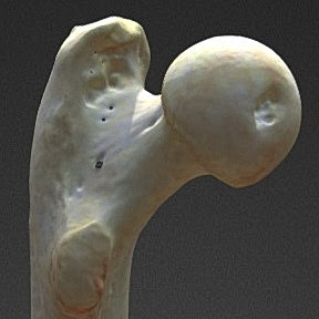
Sometimes the distal attachment site is the
edge of the head of the femur.
 |
| Orangutan. Proximal section of the right femur (posterior view); 3-D model of the femur of an adult male Bornean orangutan from the Barcelona Zoo. [sketchfab.com] |
 |
| Gorilla. Left pelvic bone and proximal section of the femur (abduction, supination); exhibit of the Zoological Museum of Moscow State University, photo by the author. |
References
Cohen KM, Harper DAT, Gibbard PL, Car N. The International Commission on Stratigraphy (ICS) International Chronostratigraphic Chart. September 2023. [stratigraphy.org]
Pyron RA. Divergence time estimation using fossils as terminal taxa and the origins of Lissamphibia. Systematic biology. 2011;60(4)466-81. [academic.oup.com]
Laurin M, Reisz RR. A reevaluation of early amniote phylogeny. Zoological Journal of the Linnean Society. 1995;113(2)165-223. [academia.edu]
Lundberg JG. Chordata. The Tree of Life Web Project. Version 1 January 1995 (under construction). [tolweb.org]
Mann A, Gee BM, Pardo JD, Marjanović D, Adams GR, Calthorpe AS, Maddin HC, Anderson JS. Reassessment of historic ‘microsaurs’ from Joggins, Nova Scotia, reveals hidden diversity in the earliest amniote ecosystem. Papers in Palaeontology. 2020;6(4)605-25. [researchgate.net]
Hedges SB, Blair JE, Venturi ML, Shoe JL. A molecular timescale of eukaryote evolution and the rise of complex multicellular life. BMC evolutionary biology. 2004;4(1)1-9. [link.springer.com]
Vaughan TA, Ryan JM, Czaplewski NJ. Mammalogy. Sudbury: Jones & Bartlett Learning, 2013. [books.google]
Meng J, Wang Y, Li C. Transitional mammalian middle ear from a new Cretaceous Jehol eutriconodont. Nature. 2011;472(7342)181-5. [researchgate.net]
Janecka JE, Miller W, Pringle TH, Wiens F, Zitzmann A, Helgen KM, Springer MS, Murphy WJ. Molecular and genomic data identify the closest living relative of primates. Science. 2007;318(5851)792-4. [epository.si.edu]
Esselstyn JA, Oliveros CH, Swanson MT, Faircloth BC. Investigating difficult nodes in the placental mammal tree with expanded taxon sampling and thousands of ultraconserved elements. Genome Biology and Evolution. 2017;9(9)2308-21. [scholar.google]
Chester SG, Bloch JI, Boyer DM, Clemens WA. Oldest known euarchontan tarsals and affinities of Paleocene Purgatorius to Primates. Proceedings of the National Academy of Sciences. 2015;112(5)1487-92. [scholar.google]
Smith T, Rose KD, Gingerich PD. Rapid Asia-Europe-North America geographic dispersal of earliest Eocene primate Teilhardina during the Paleocene-Eocene Thermal Maximum. Proc Natl Acad Sci USA. 2006;103:11223-7. [scholar.google]
Srivastava RP. Morphology of the Primates and Human Evolution. New Delhi: PHI Learning Pvt. Ltd., 2009. [books.google]
Gebo DL, MacLatchy L, Kityo R, Deino A, Kingston J, Pilbeam D. A hominoid genus from the early Miocene of Uganda. Science. 1997;276:401-4. [researchgate.net]
Leakey M, Walker A. Afropithecus: function and phylogeny. In: Begun DR, Ward CV, Rose MD (Eds). Function, phylogeny and fossils: Miocene hominoid evolution and adaptations. New York: Plenum, 1997:225-39. [link.springer.com]
Israfil H, Zehr SM, Mootnick AR, Ruvolo M, Steiper ME. Unresolved molecular phylogenies of gibbons and siamangs (Family: Hylobatidae) based on mitochondrial, Y-linked, and X-linked loci indicate a rapid Miocene radiation or sudden vicariance event. Molecular Phylogenetics and Evolution. 2011;58(3)447-55. [ncbi.nlm.nih.gov]
Poszewiecka B, Gogolewski K, Stankiewicz P, Gambin A. Revised time estimation of the ancestral human chromosome 2 fusion. BMC genomics. 2022;23(6)1-16. [link.springer.com]
Наумов НП, Карташев НН. Зоология позвоночных. Ч. 2. Пресмыкающиеся, птицы, млекопитающие: Учебник для биолог. спец. ун-тов. Москва: Высшая школа, 1979. [chembaby.ru]
Wilson DE, Reeder DM (Eds). Mammal species of the world: a taxonomic and geographic reference. Vol. 1. Baltimore: Johns Hopkins University Press, 2005. [books.google]
Groves CP. Towards a taxonomy of the Hominidae. In: Humanity from African Naissance to Coming Millennia. In: Tobias PV, Raath MA, Moggi-Cecchi J, Doyle GA (Eds). Colloquia in Human biology and Palaeonthropology. Firenze: Firenze University Press, 2001:291-7. [library.oapen.org]
Crelin ES. Ligament of the head of the femur in the orangutan and indian elephant. The Yale J Biol Med. 1988;61(5)383-8. [ncbi.nlm.nih.gov, ncbi.nlm.nih.gov.pdf]
Owen R. On the osteology of the Chimpanzee and Orang. Transactions of the Zoological Society of London. Vol. I. London, 1835:343-379. [books.google]
Mivart G. Contributions towards a more complete knowledge of the Skeleton of the Primates. Part I. The Appendicular Skeleton of Simia. Transact. Zool. Soc. 1869;6:175-226. [biodiversitylibrary.org]
Moser E. Ueber das Ligamentum teres des Hüftgelenks. Morphologische Arbeiten. 1893;2(1)36-92. [books.google , jstor.org]
Keywords
ligamentum capitis femoris, ligamentum teres, ligament of head of femur, doctrine, animals, monkey, homo
The original text in Russian is available at the link: Primates
NB! Fair practice / use: copied for the purposes of criticism, review, comment, research and private study in accordance with Copyright Laws of the US: 17 U.S.C. §107; Copyright Law of the EU: Dir. 2001/29/EC, art.5/3a,d; Copyright Law of the RU: ГК РФ ст.1274/1.1-2,7
Comments
Post a Comment