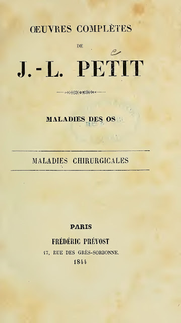
1844PetitJL


Fragments from the book Petit JL. Maladies des os, maladies chirurgicales (1844). Reprint of an 18th-century author's work discussing the anatomy, role, and damage of the ligamentum capitis femoris (LCF) in hip dislocation. The text is prepared for machine translation using a service built into the blog from Google or your web browser. 

Quote p. 80

DE LA LUXATION DE LA CUISSE.

La tête du fémur est très-grosse, et la cavité de l'ischion fort profonde. Elles sont l'une et l'autre revêtues d'un cartilage poli, excepté cependant aux endroits où il n'y a point de frottement, c'est-à-dire aux attaches d'un ligament qui se trouve dans l'intérieur de l'article, et qui s'insère à la tête du fémur, un peu au-dessous du milieu, et prend origine de la partie excentrale et inférieure de la cavité cotyloïde , où se trouve un enfoncement pour loger les glandes synoviales.

La cavité cotyloïde est plus profonde en haut et en arrière qu'en bas et en devant. Elle a, par sa partie inférieure, une échancrure fermée par un ligament sous lequel passent les vaisseaux qui portent la nourriture au ligament intérieur connu sous le nom de ligament rond, aux glandes synoviales et aux autres parties de l'article. 

Quote p. 81

4. Le ligament rond s'oppose à l'éloignement de l'os, non à la vérité dans tous les sens, parce qu'il n'est pas attaché précisément dans le plus profond de la cavité ni au milieu de la tête, mais du moins, comme on va le faire observer, résiste-t-il à plusieurs espèces de luxations.

Des différentes Espèces de Luxations de la Cuisse.

2. Le ligament rond se trouvant plus proche du bord de la cavité du côté interne, la tête de l'os peut s'éloigner plus de ce côté que des autres sans que le ligament s'y oppose.

Par des raisons contraires la luxation doit arriver plus rarement en haut. 1° Les bords de la cavité y sont plus élevés. — 2° L'os ne peut être luxé de ce côté que le ligament rond ne soit rompu, et que par conséquent l'effort ne soit très-violent; car, s'il était médiocre, ce ligament , capable d'une certaine résistance , pourrait empêcher l'éloignement de la tête de l'os. — 3° Enfin les muscles les plus puissans s'opposent à cette luxation. 

Quote p. 84

Du Pronostic de la Luaation de la Cuisse.

Lorsque la cuisse est luxée en haut, la guérison est difficile et incertaine, quoique la réduction ait été bien faite, ce qui vient de ce que le ligament rond souffre nécessairement rupture dans ces sortes de luxations, et de ce que la réunion ne s'en fait pas toujours, quelque précaution qu'on prenne pour la procurer. 

Quote p. 87

De la Cure de la Luxation de la Cuisse.

Les luxations en haut exigent qu'on applique des appareils plus serrés , et qu'on fasse garder le repos beaucoup plus exactement qu'après les autres luxations ; et cela à cause de la rupture du ligament rond, dont la réunion se fait difficilement, et demande un temps considerable. 

Quote p. 91

De la Luxation de la Cuisse qui succède auxchutes du grand Trochanler.

Lorsque, dans une chute, le grand trochanter est frappé, la tête du fémur est violemment poussée contre les parois de la cavité cotyloïde ; et , comme elle remplit exactement cette cavité , les cartilages , les glandes de la synovie et le ligament de l'intérieur de l'article doivent souffrir une forte contusion, qui sera suivie d'obstruction, d'inflammation et de dépôt. La synovie surtout s'amassera dans la cavité de l'articulation; la capsule ou tunique ligamenteuse en sera distendue, et la tête de l'os, peu à peu chassée au dehors, sera enfin entièrement luxée.

 '''La synovie s'épanchant continuellement dans l'article, s'y épanchant même alors plus que dans l'état naturel, et n'étant plus dissipée par les mouvemens de la partie, on ne doit point être surpris qu'elle s'accumule, et qu'elle remplisse la cavité au point de chasser la tête de l'os ; ce qu'elle fera avec d'autant plus de facilité que, relâchant les ligamens, elle les met hors d'état de résister non-seulement à la force avec laquelle elle pousse l'os hors de sa boîte, mais encore aux efforts que font les muscles pour tirer en haut la tête du femur. La capsule ne sera donc pas seule distendue, le ligament rond souffrira aussi peu à peu un alongement qui sera accompagné d'une douleur trèsvive, laquelle augmentera par degrés, et ne diminuera que quand ce ligament, tout-à-fait relâché ou rompu, aura abandonné la tête de l'os à toute la puissance des muscles qui la tirent en. 

Quote p. 92

Si la tête de l'os, entièrement chassée de sa cavité, n'est pas d'abord portée plus loin par l'action des muscles, c'estparcequele ligament rond la retient encore; et il est facile de concevoir qu'alors les douleurs doivent augmenter considérablement. En effet, tant que quelque portion de la tête a pu être retenue par le rebord de la cavité yle ligament rond a partagé avec lui l'effort des muscles, et ne-s^.est alongé que peu à peu; mais, la tête du fémur ayant été entièrement chassée, le ligament supporte lui seul l'effort des muscles, les douleurs deviennent insupportables, et durent, comme on l'a déjà dit, jusqu'à ce que la rupture du ligament ou sa relaxation entière ait permis aux muscles d'éloigner l'os autant qu'il peut l'être parleur plus grande contraction. 

External links

Petit JL. Oeuvres completes de J.L. Petit: Maladies des os, maladies chirurgicales. Paris: Frederic Prevost, 1844. [archive.org]

Authors & Affiliations

Jean-Louis Petit (1674-1750) was French surgeon and anatomist. [wikipedia.org]

Jean-Louis Petit
The author of the image is Ambroise Tardieu;
original in the 
wikimedia.org collection (CC0 – Public Domain, no change)

Keywords

ligamentum capitis femoris, ligamentum teres, ligament of head of femur, anatomy, dislocation, injury, pathological anatomy


NB! Fair practice / use: copied for the purposes of criticism, review, comment, research and private study in accordance with Copyright Laws of the US: 17 U.S.C. §107; Copyright Law of the EU: Dir. 2001/29/EC, art.5/3a,d; Copyright Law of the RU: ГК РФ ст.1274/1.1-2,7

DISEASES AND INJURIES

Comments

Popular posts from this blog

4cent.Gothic Bible

  About the Book of Genesis of the Gothic Bible (4th century). A Gothic term that may have been used to refer to the ligamentum capitis femoris (LCF) of an animal and a human. See our commentary at the link: 4cent.Gothic Bible [Rus]. Quote [ Got ] Genesis 32:33 (possible term;  original source  lost) Translation [Eng] Genesis 32:33 The original text and English translation are currently unavailable to our project. Suggested term: … gabinda ( gabundi) … The closest analogue is in the Epistle to the Colossians 2:19 — jah ni habands haubiþ, us þammei all leik þairh gawissins jah gabindos auknando jah þeihando wahseiþ du wahstau gudis. — καὶ οὐ κρατῶν τὴν κεφαλήν , ἐξ οὗ πᾶν τὸ σῶμα διὰ τῶν ἁφῶν καὶ συνδέσμων ἐπιχορηγούμενον καὶ συμβιβαζόμενον αὔξει τὴν αὔξησιν τοῦ θεοῦ . — And not holding the Head, from which all the body by joints and bands having nourishment ministered, and knit together, increaseth with the increase of God. (original source: wulfila.be ; Codex Ambrosianus? Milan, Bi

2018FreitasA_BandeiraVC

  The authors reported a case of open hip dislocation and documented avulsion of the ligamentum capitis femoris (LCF) from the acetabulum.  The injured LCF appeared as a large cord-like structure, attached to the femoral head ( Fig. 1 ). Perhaps Hippocrates of Kos saw such an LCF in open dislocation of the hip and described it in his treatise «Mochlicus» ( 1886AdamsF ). One year later, the patient's x-ray revealed a significant narrowing of the joint space in the upper part of the hip joint ( Fig. 9 ). In our opinion, this to be a consequence of hyaline cartilage wear due to overload in the absence of LCF. In an unaffected hip joint, the width of the radiological joint space at the upper section than in the lower section ( Ossification of the LCF ). When examined one year after the injury, the patient could stand on one leg. We noticed that his body was excessively deviated towards the supporting hip joint, and the opposite half of the pelvis was elevated ( Fig. 8 ). This is a me

1875BrauneW

  We present a study dedicated to the experimental investigation of the function of the ligamentum capitis femoris (LCF). Wilhelm Braune established that the LCF prevents hip supination and adduction, especially during flexion. In the conclusion of the study, the author writes: "It also remains to determine in which body movements this ligament function plays a role, as it is currently unclear why the femur requires a special fixing device…". Apparently, Wilhelm Braune did not have time to find the answer, since his outstanding monograph on LCF does not say a word ( Braune W, Fischer O. Der Gang des Menschen (1895 books.google ) . Based on our own experiments on mechanical models and research, we believe that adduction and supination of the hip, along with flexion, are observed at the beginning of the single-support period of the step. At this moment, tension occurs in the LCF, allowing to reduce the load on the upper segment of the femoral head. The LCF is also stretched in

927-942Arabic Bible

  Fragment of the Book of Bereshit   translated into Arabic by Saadia Gaon  ( 927-942 ) . The Arabic text contains mentioned to ligamentum capitis femoris (LCF) of an animal and a human. See our commentary at the link: 927-942ArabicBible [Rus].  Quote [ a ra] التكوين  32:32 (original source: 1653WaltonB, p. 145) See also modern editions:   لذلك لا يأكل بنو اسرائيل عرق النّسا الذي على حقّ الفخذ الى هذا اليوم . لانه ضرب حقّ فخذ يعقوب على عرق النّسا (original source: arabicbible.com )  لِذَلِكَ لا يَاكُلُ بَنُو اسْرَائِيلَ عِرْقَ النَّسَا الَّذِي عَلَى حُقِّ الْفَخِْذِ الَى هَذَا الْيَوْمِ لانَّهُ ضَرَبَ حُقَّ فَخْذِ يَعْقُوبَ عَلَى عِرْقِ النَّسَا (original source: copticchurch.net ) Translation [Eng] Genesis 32:32 Translation of the text is currently unavailable for our project. The term for the LCF:  النّسا   ~  sciatica  ( see also: 70-110Rufus Ephesius ; 180-238PolluxJ ) Saʻadia ben Joseph . Pentateuch ( 1600) , p. 1  ( original:  digital.library.yu.edu ) External links Saʻadia

2016ArkhipovaAS Classification of Joints with Flexible Elements.

  Classification of Joints with Flexible Elements The classification of joints with flexible elements was first proposed by A.S. Arkhipova in 2016 and presented at conferences in 2016-2017 ( 2017АрхиповаАС ). Joints with flexible elements are divided by the author into detachable and non-detachable, three-axis, two-axis, and single-axis. Each can contain in various combinations one or more external and internal flexible elements - analogs of joint ligaments, including the ligamentum capitis femoris (LCF). Conditional designations of joints with flexible elements using the example of a ball joint: Detachable ball joint with an internal flexible element Non-detachable ball joint with an internal flexible element Detachable ball joint with an external flexible element Non-detachable ball joint with an external flexible element Detachable ball joint with both internal and external flexible elements Non-detachable ball joint with both internal and external flexible elements Flexible e

1879MorrisH

  Fragments of the book Morris H. The anatomy of the joints of man (1879) dedicated to ligamentum capitis femoris (LCF). The author discusses the anatomy of the LCF and describes his experiments to study its movement.   Quote p p . 318-319 The acetabulum is partly articular, partly non-articular. The articular portion is of horseshoe shape, and extends inwards from the margin, more or less. It is altogether deficient at the cotyloid notch, which corresponds with the gap of the horseshoe. It is the widest at the iliac part, where it is over one inch from without inwards; then it very gradually gets narrower along the ischium, but widens out again at the ischial end of the cotyloid notch ; forwards from the pubo-iliac suture it narrows more rapidly, and does not extend quite up to the pubic end of the cotyloid notch. At its narrowest point in an adult bone it measures half an inch in width. When coated with cartilage, and fringed round with the cotyloid ligament, it fits very closel

DIAGNOSTICS AND EXAMINATION

  DIAGNOSTICS AND EXAMINATION   (Diagnostic, examination and testing methods... ) Catalog. LCF Pathology Tests   Tests for the detection of pathology LCF.  2004VialleR_GlorionC  The article discusses the examination technique for dislocation of the femur and describes the radiographic symptom of infringement of the damaged LCF. BLOG CONTENT

150-250Targum Jonathan

  Fragments from the Targum Jonathan on Genesis. Tractate was written between about 150 - 250 in lend of Israel. The text is a combination of a translation and commentary on the book of Bereshit. The unknown compiler mentions ligamentum capitis femoris (LCF) in an animal and an episode of its damage in a human. See our commentary at the link: 150-250Targum Jonathan [Rus]. Quote 1. [Heb] Genesis. 32:33 (original source:  sefaria.org ) Quote 2. [Heb] Genesis. 43:16 (original source:  sefaria.org ) Translation Quote 1. [Eng] Genesis. 32:33 Therefore, the sons of Israel eat not the sinew which shrank, which is in the hollow of the thigh of cattle and of wild animals, until this day; because the Angel touched and laid hold of the hollow of the right thigh of Jakob, in the place of the sinew which shrank. (Transl. by J.W. Etheridge (186 2 ) ; original source: targum.info ) Quote 2. [Eng] Genesis. 43:16 And Joseph saw Benjamin with them: and he said to Menasheh whom he had made superintende

1679DiemerbroeckI

  Fragments from the book Diemerbroeck I. Anatome corporis humani (1679). The author describes the pathology variants, function, topography and attachment of the ligamentum capitis femoris (LCF). The damage to the LCF in hip dislocation, symptoms and treatment are discussed. The text is similar to a paraphrase of Hegetor's work «On Causes» and Galen of Pergamon's commentary on Hippocrates' treatise «On Joints» ( 1745CocchiA ;  2020ArkhipovSV_ProlyginaIV ). Isbrand van Diemerbroeck uses many synonyms for LCF: nervus cartilaginosus, terete, teres, rotundum ligamentum, interius ligamentum. Quote p. 593. [Lat] CAPUT XIX. De Ossibus Femoris, & Cruris. Superius procesum crassum, versus coxendicis os prominentem, eique epiphyin rotundam & amplam impositam habet, sicque globosum femoris caput, valida cervice subnixum, constituit, quod cartilagine obductum in coxendicis acetabulum reconditur, in eoque duobus validis ligamentis detinetur: uno lato, crasso, & membranoso,

2014ArkhipovaAS

  In 2014, at the International Olympics Space for school students, Alexandra Arkhipova presented a report on the feasibility of using joints with flexible elements in walking machines. The author was recognized as the overall winner (more details: cyclowiki.org ). The report suggested: «An important area of possible application of walking robots would be remote exploration of other planets». Ten years later, professionals from NASA began to put this idea into practice: Robot dog trains to walk on Moon in Oregon trials (more details: bbc.com ). Below we present the text of the first message about walking machines in space, the ball joint of which contains a flexible element - an analogue of the ligamentum capitis femoris (LCF).   Mobile biomorphic platforms with analogues of natural locomotion algorithms Arkhipova A.S. For ten years of its mission, American wheeled Mars rover vehicle 'Opportunity' covered just 40 kilometers through the Red Planet, and its twin Spirit go