
1875HeitzmannC


Fragments from the book Heitzmann C. Die descriptive und topographische Anatomie des Menschen in 600 Abbildungen (1875). The author briefly describes the topography and blood supply of the ligamentum capitis femoris (LCF), and also supplements the text with original illustrations.

The text is prepared for machine translation using a service built into the blog from Google or your web browser.

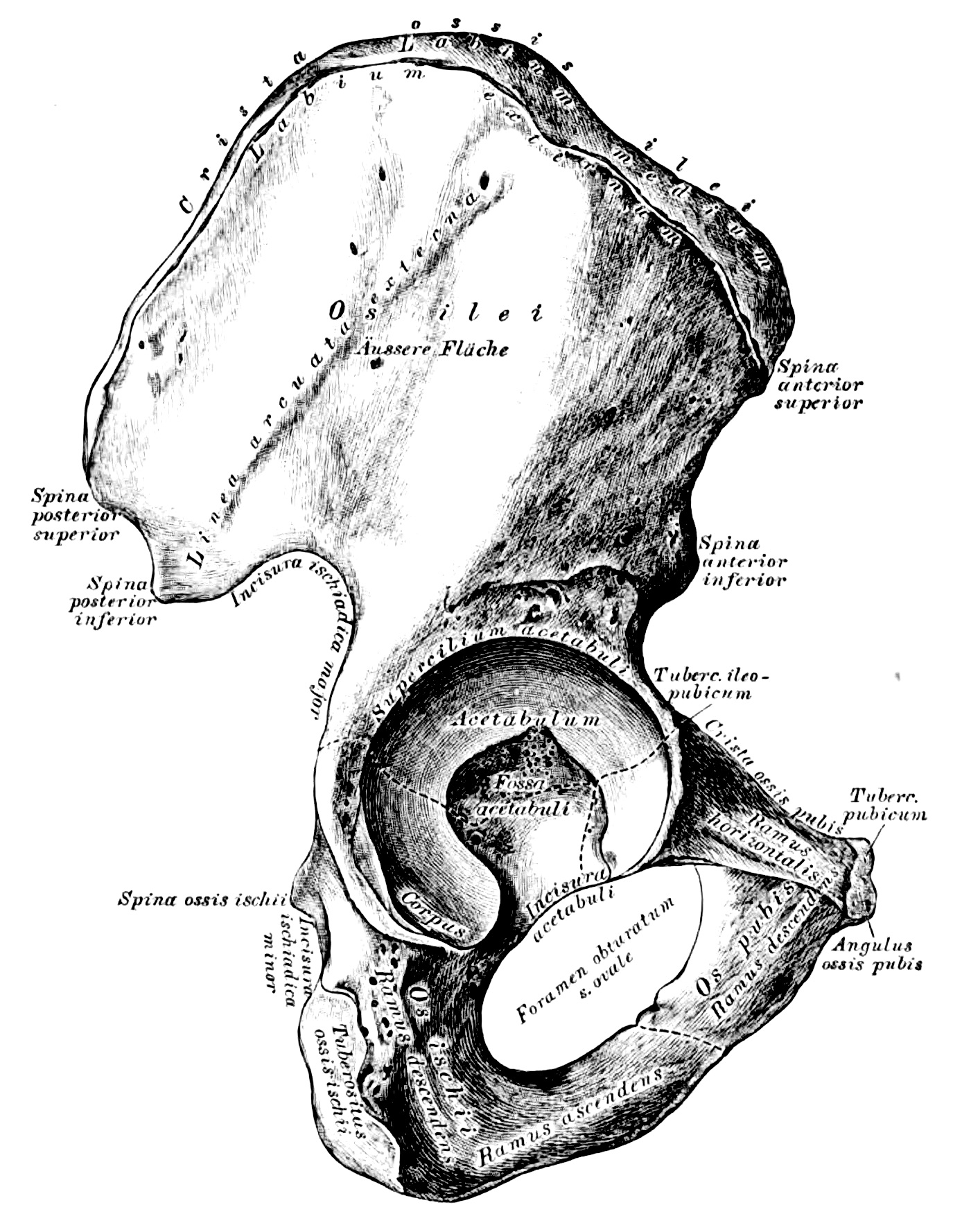
150. Das rechte Hüftbein, Os innominatum, von aussen.

153. Das rechte Hüftbein, Os innominatum, von innen. mit den Muskelansätzen.

165. Das rechte Oberschenkelbein, Os femoris, von vorne [fragment].

Quote pp. 119-123 

Am oberen Ende des Oberschenkelbeines fällt der an einem Halse sitzende Kopf, Caput femoris in die Augen, mit einem Grübchen, Foveola, zur Anheftung des Lig. teres. An der Uebergangsstelle des Halses in das Mittelstück stehen die beiden Rollhügel, der grosse äussere, Trochanter major, und der kleine innere, Trochanter minor; dieselben sind durch die Linea intertrochanterica anterior und posterior vereinigt. Nach innen vom grossen Trochanter liegt die Fossa trochanterica (Fig. 166). An der hinteren Fläche des Mittelstückes springt die in 2 Lefzen (Labia) gespaltene Linea aspera femoris vor (F. 166).

Das untere Ende ist mit 2 Knorren, Condylus externus und Condylus internus, versehen, deren jeder eine Tuberosität besitzt. Die Knorren sind an der hinteren Seite durch die Fossa poplitea von einander getrennt. (Fig. 166).

Das Oberschenkelbein ist mit 3 Knochen gelenkig verbunden: mit dem Hüftbeine, dem Schienbeine, und der Kniescheibe

166. Das rechte Oberschenkelbein, Os femoris, von hinten [fragment, left].
167. Das rechte Oberschenkelbein, Os femoris,von hinten, mit den Muskelansätzen [fragment, right].


168. Das rechte Hüftgelenk, Articulatio coxae, von vorne.

Die fibröse Kapsel des Hüftgelenkes inserirt am Umfange des knöchernen Pfannenrandes einerseits, und an der Vorderseite des Oberschenkelknochens an der Linea intertrochanterica anterior andererseits. Als Verstärkung der vorderen Kapselwand dient das kräftige, von der Spina anterior inferior des Darmbeines entspringende Ligamentum Bertini, welches zum Theile zur Linea intertrochanterica anterior herabsteigt (Fig 154), zum Theile mit 2 Schenkeln den Oberschenkelhals als Zona orbicularis Weberi umschlingt (Fig. 156).

169. Das rechte Hüftgelenk, Articulatio coxae, eröffnet.

Am knöchernen Umfange der Pfanne haftet ringsum ein faserknorpeliger Ring, Limbus cartilagineus acetabuli; an der Stelle der Incisura acetabuli bildet dieser Ring eine Brücke. Von der Foveola des Oberschenkelkopfes zieht zur nicht überknorpelten Fovea acetabuli das runde Band, Ligamentum teres. Die fibröse Kapsel des Hüftgelenkes ist in der Figur aufgeschnitten und zurückgelegt dargestellt; es wird ersichtlich, dass an der vorderen Seite der Schenkelhals vollständig von der Kapsel eingehüllt ist.

170. Das rechte Hüftgelenk, Articulatio coxae, im Durchschnitte.

Die fibröse Kapsel, an der vorderen Seite des Gelenkes sehr stark, ist am hinteren Umfange desselben weit schwächer, und haftet nicht an der Linea intertrochanterica posterior, sondern indem sie sich umbiegt, an der hinteren Fläche des Schenkelhalses. Das Ligamentum teres steigt von der Incisura acetabuli zur Foveola des Oberschenkelkopfes senkrecht hinauf; dasselbe ist von der Synovialkapsel eingehüllt. Die Abbildung zeigt auch die eigenthümlich angeordnete Knochenstructur des Oberschenkelhalses und des Kopfes.

Quote p. 215

Die vorderen Aeste der 4. hypogastrica sind:

a) Die A. obturatoria geht in Begleitung des N. obturatorius durch den Canalis obturatorius, und zerfällt am oberen Rande des M. obturatorius in einen Ramus anterior und einen posterior. Der erstere verästelt sich im M. adductor femoris longus et brevis, pectineus und gracilis; der letztere sendet die A. acetabuli zum Lig. teres des Oberschenkelkopfes und löst sich schliesslich in den Auswärtsrollern auf. 


External links

Heitzmann C. Die descriptive und topographische Anatomie des Menschen in 600 Abbildungen. Erster band: 1. Knochen, Gelenke, Bander. 2. Muskeln, Fascien, Topographie. 3. Sinneswerkzeuge. Wien: W. Braumuller, 1875. [books.google]

Heitzmann C. Die descriptive und topographische Anatomie des Menschen in 600 Abbildungen. Erster band: 1. Knochen, Gelenke, Bander. 2. Muskeln, Fascien, Topographie. 3. Sinneswerkzeuge. Wien: W. Braumuller, 1870.  [archive.org]

Heitzmann C. Die descriptive und topographische Anatomie des Menschen in 637 Abbildungen Fünfte auflage. Wien: W. Braumuller, 1888.  [books.google]

Authors & Affiliations

Carl Heitzmann (1836-1896) was a pathologist, dermatologist in the Austro-Hungarian Empire and United States of America. [wikipedia.org] 

Carl Heitzmann 
Unknown date and author; 
original in the enciklopedija.hr collection
(CC0 – Public Domain, no changes)

Keywords

ligamentum capitis femoris, ligamentum teres, ligament of head of femur, anatomy, topography, vascularization


NB! Fair practice / use: copied for the purposes of criticism, review, comment, research and private study in accordance with Copyright Laws of the US: 17 U.S.C. §107; Copyright Law of the EU: Dir. 2001/29/EC, art.5/3a,d; Copyright Law of the RU: ГК РФ ст.1274/1.1-2,7

MORPHOLOGY AND TOPOGRAPHY

Comments

Popular posts from this blog

1981OrtnerDJ_PutscharWGJ

  Content [i] Annotation [ii] Original text [iii] Illustrations [iv] Source & links [v] Notes [vi] Authors & Affiliations [vii] Keywords [i] Annotation Fragment from the book: Ortner DJ, Putschar WGJ. Identification of Pathological Conditions in Human Skeletal Remains (1981). The authors describe signs of ligamentum capitis femoris (LCF) pathology in the remains of a Bronze Age individual, possibly resulting from recurrent hip subluxation. The text in Russian is available at the following link: 1981OrtnerDJ_PutscharWGJ . [ii] Original text Quote pp. 360-361   An adult male skeleton from tomb A100E at the Early Bronze Age [c. 3500–2000 BCE] cemetery of Bab edh-Dhra in Jordan has an abnormally shallow acetabulum of the right hip. This is the same skeleton that had a separate neural arch. The acetabulum is also much larger in diameter than the corresponding acetabulum of the left innominate (Figure 577). There is a moderate amount of arthritic lipping on the m...

1845HollsteinL

  Fragments from the book Hollstein L. Compendium der Anatomie des Menschen (1865). The author discusses the anatomy of the ligamentum capitis femoris (LCF), and mentions its synonyms. The text is prepared for machine translation using a service built into the blog from Google or your web browser.   Quote p. 144 Das Lig am. teres s. rotundum femoris (Fig. 50, 7.) hat eine dreieckige Gestalt, und ist mit seiner Basis in dem Fundus acetabuli, mit seiner rundlichen Spitze in der Fovea capitis ossis femoris befestigt. Es besteht aus einem Bündel Sehnenfasern, und erhält von der Synovialmembran einen scheidenformigen Ueberzug; bisweilen existirt letzterer allein, und manchmal felilt das Band ganz und gar. Fig. 50. Seitliche Ansicht der Bänder des Beckens und Hüftgelenks.   Quote p. 145 Die weite Synovialmembran überzieht den Schenkelkopf, geht alsdann als Scheide des Ligam. teres zur Gelenkpfanne über, welche sie ebenfalls auskleidet, und schlägt sich hierauf über die ...

Vertebrates

VERTEBRATES According to the molecular clock, a specific method for dating phylogenetic events, vertebrates (Vertebrata) separated from arthropods (Arthropoda) 976±97 Ma (2004HedgesSB_ShoeJL). The latter began to dominate in species diversity with the Cambrian burst of radiation, which occurred 520 Ma (2010EdgecombeGD). This ratio in the fauna of the Earth is still preserved. Approximately 525 Ma, the phylum Chordates separated from the group of bilaterally symmetrical animals (1995ChenJY_ZhouGQ). In turn, the evolution of chordate organisms led to the formation of the first vertebrates at least 500 Ma, from which the jawed mouths 450-400 Ma descended, becoming the ancestors of the placoderms or "armored" fish (Placodermi) (1979 НаумовНП _ КарташевНН ). Sculptural reconstruction of the placoderm Coccosteus from the order Arthrodires, Middle Devonian, 393.3-382.7  Ma ; exposition of the Orlov Paleontological Museum (Moscow); photo by the author. The first cartilaginou...

Online Journal «ABOUT ROUND LIGAMENT OF FEMUR», May 2026

  The journal is dedicated to the  ligamentum capitis femoris (LCF)  and related topics   About the Journal »»»                                                                                . The online journal « About Round Ligament of Femur » was created based on the scientific blog of the same name. The resource is the English-language part of the project:  ONLINE JOURNAL: Ligamentum capitis femoris .   Updates: As new materials are prepared. Mission : Popularization and preservation of knowledge about LCF, as well as promoting its practical application. Main goal: Improvement of diagnosis, treatment, and prevention of injuries and diseases of the hip joint. Publisher: Arkhipov S.V., independent researcher, PhD, orthopedic surgeon. Reviewers: G...

1803LarreyDJ

  Content [i] Annotation [ii] Original text [iii] English translation [iv] Source & links [v] Notes [vi] Authors & Affiliations [vii] Keywords [i] Annotation Fragment from the book: Larrey DJ. Relation historique et chirurgique de l'expedition de l'armée d'Orient, en Egypte et Syrie (Historical account and surgery of the expedition of the Army of the Orient, in Egypt and Syria, 1803). The author describes exarticulation in the hip joint and the technique of cutting the ligamentum capitis femoris (LCF), which he calls the "interarticular ligament". The text in Russian is available at the following link: 1803LarreyDJ . [ii] Original text (France ) Quote pp. 325-328   Les praticiens qui ont proposé l'extirpation de la cuisse ne sont point d'accord sur la manière de la faire; cependant presque tous, craignant l'hémorragie de l'artère crurale, commencent par la ligature de ce vaisseau, forment ensuite un lambeau aux dépens des muscles ...

1948EpsteinI

  Content [i] Annotation [ii] Original text [iii] Illustration [iv] Source & links [v] Notes [vi] Authors & Affiliations [vii] Keywords [i] Annotation Fragments from the book: Epstein I. Babylonian Talmud. Seder Kodashim. Vol. 2. Hullin (1948). The editor comments on the words of Rabbi Samuel (Shmuel) in the tractate Hullin of the Babylonian Talmud, explaining the location of the ligamentum capitis femoris (LCF) in relation to the joint and its differences from the sciatic nerve. The original in Russian is available at: 1948EpsteinI . [ii] Original text Quote p. 500 Hullin. Chapter 7.89b MISHNAH. [THE PROHIBITION OF] THE SCIATIC NERVE(1) IS IN FORCE BOTH WITHIN THE HOLY LAND AND OUTSIDE IT, BOTH DURING THE EXISTENCE OF THE TEMPLE AND AFTER IT, IN RESPECT OP BOTH UNCONSECRATED AND CONSECRATED [ANIMALS]. IT APPLIES TO CATTLE AND TO WILD ANIMALS, TO THE RIGHT AND LEFT HIP, BUT IT DOES NOT APPLY TO BIRDS BECAUSE THEY HAVE NO SPOON-SHAPED HIP(2). IT ALSO APPLIES TO A F...

1665LindenJA

  Content [i] Annotation [ii] Original text (in Latin) [iii] English translation [iv] Source & links [v] Notes [vi] Authors & Affiliations [vii] Keywords [i] Annotation Fragment from the book: Linden JA . Magni Hippocratis Coi Opera Omnia Graece Et Latine Edita. Vol. I. (1665). This article presents an excerpt from the treatise «Mochlicus» (Instruments of Reductions) by  Hippocrates of Cos    (b. 460 BC), translated into Latin. The author describes for the first time the localization and area of distal attachment of the ligamentum capitis femoris (LCF) describit, mentionem in alio tractatu ponens. A translation of this article into Russian is available at the link: 1665LindenJA .  The original source in Greek sees at the link: 1844LittreE , and in English at: 1886AdamsF . [ii] Original text (in Latin) Quote pp. 294-295 Vol. I. Ossium natura II. Ipsum aurem femur foras, & in anteriore parte incurvum est. Caput autem ejus appendix eft r...

THE DOCTRINE OF LCF

  THE DOCTRINE OF  ligamentum capitis femoris:   An Instrument of Knowledge and Innovation. Definition: A set of theoretical provisions on all aspects of knowledge about the anatomical element ligamentum capitis femoris (LCF). 1. Structure of the Doctrine of LCF 2.  Practical Application of the Doctrine of LCF : 2.1. Diagnostics 2.1. Prevention   2.3. Prognosis 2.4. Pathology 2.5. Veterinary   2.6. Professions     2.7. Products     2.8. Surgery   3. Theory of LCF Mechanics    4. The Base of the Doctrine of LCF 5. Stairway to the Past or History of the Doctrine of LCF 6. Ultimate Depth of Researches   7. Appendices 7.1. Acceptable Synonyms      Structure of the Doctrine of  ligamentum  capitis  femoris .       E     a     R                   T                   ...

Who, When, and Where Wrote the Book of Genesis?

  Who, When, and Where Wrote the Book of Genesis?  A Medical Hypothesis By Sergey V. Arkhipov, MD, PhD & Lyudmila N. Arkhipova, BSN     CONTENT [i]   Abstract [ii]   Introduction [iii]   Egyptian physician [iv]   Asian diviner [v]   Conclusion [vi]   References [vii]   Application [i]   Abstract The Book of Genesis is an example of an ancient literary text that contains important medical insights. We propose that it was written in northern Egypt in the late 17th century BCE, approximately ten years after the Minoan eruption. The protograph likely emerged from the collaboration between an Asiatic seer, who rose to the rank of an Egyptian official, and an Egyptian physician-encyclopedist. By refining its dating and authorship, this hypothesis positions Genesis as a credible source of medical and historical data, thereby enhancing its value for interdisciplinary research. [ii]   Introduction According to Rabbinic Judais...

1906HartmannO

  Hartmann O. s ketch of a painting , Jacob wrestling with the angel (1906?).   Depicting the circumstances and mechanism of the ligamentum capitis femoris (LCF) injury based on the description in the Book of Genesis:  25 And Jacob was left alone; and there wrestled a man with him until the breaking of the day. 26 And when he saw that he could not pre vail against him, he struck against the hollow of his thigh ; and the hollow of Jacob's thigh was put out of joint, as he was wrestling with him. … 33 Therefore do the children of Israel not eat the sinew which shrank, which is upon the hollow of the thigh, unto this day; because he struck against the hollow of Jacob's thigh on the sinew that shrank.  ( 1922LeeserI , Genesis (Bereshit) 32:25-26,33) More about the plot in our work:  Ninth month, eleventh day   ( 2024 АрхиповСВ. Девятый месяц, одиннадцатый день ).     Oluf Hartmann  – Jacob Wrestling with the Angel (1906?); original in the  ...