2016ArkhipovaAS Classification of Joints with Flexible Elements.


Classification of Joints with Flexible Elements

The classification of joints with flexible elements was first proposed by A.S. Arkhipova in 2016 and presented at conferences in 2016-2017 (2017АрхиповаАС).

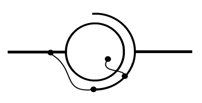
Joints with flexible elements are divided by the author into detachable and non-detachable, three-axis, two-axis, and single-axis. Each can contain in various combinations one or more external and internal flexible elements - analogs of joint ligaments, including the ligamentum capitis femoris (LCF).

Conditional designations of joints with flexible elements using the example of a ball joint:


Detachable ball joint with an internal flexible element


Non-detachable ball joint with an internal flexible element

Detachable ball joint with an external flexible element

Non-detachable ball joint with an external flexible element

Detachable ball joint with both internal and external flexible elements


Non-detachable ball joint with both internal and external flexible elements

Flexible elements can be used to secure the head in the socket, as well as to change the trajectory and range of motion of the joint. The length of the flexible elements and the location of their attachment points play an important role. Joints with flexible elements can be used inin pedipulators of walking machines, robots, and exoskeletons, as well as in endoprosthetics of human and animal joints. 

Author and Organization:

Arkhipova Alexandra Sergeevna

Currently (2024) a 6th-year student at the MSU Faculty of Mechanics and Mathematics (Механико-математический факультет Московского государственного университета имени М.В. Ломоносова)

References:

Arkhipova AS. Study of properties of joints with flexible elements for biomorphic walking technology. Research work, conducted at the Lyceum of Scientific and Engineering Profile. Korolev, 2016. [in Russian] (Архипова АС. Изучение свойств шарниров с гибкими элементами для биоморфной шагающей техники. Научно-исследовательская работа, выполненная в АОУ Лицей научно-инженерного профиля. Королёв, 2016.)

Arkhipova AS. Study of properties of joints with flexible elements for biomorphic walking technology. Presentation of the report. Moscow, 2017. [in Russian] (Архипова АС. Изучение свойств шарниров с гибкими элементами для биоморфной шагающей техники. Презентация доклада. Москва, 2017.)

Keywords:

joints with flexible elements, classification

                                                                    .

In translating to English, the author is assisted by ChatGPT (version 3.5) and the Google Translate service.

BLOG CONTENT 

REVIEWS AND CLASSIFICATIONS

Comments

Popular posts from this blog

LCF in 2026 (September)

  LCF in 2026 (September )  (Quotes from articles and books published in September   2026 mentioning the ligamentum capitis femoris)   Goetz, J. E., Hockman, J., & Willey, M. C. (2026). Rabbit models of hip dysplasia: a narrative review. Laboratory animal research , 42 (1), 32.   [i]   link.springer.com   Kahana-Rojkind, A. H., Strok, M. J., Walsh, E. G., Quesada-Jimenez, R., Kuhns, B. D., & Domb, B. G. (2026). Factors Associated With Return to Sport at Minimum 10-Year Follow-up in Athletes With Borderline Hip Dysplasia Undergoing Hip Arthroscopy With Capsular Plication. Orthopaedic Journal of Sports Medicine , 14 (9), 23259671261451218.   [ii]   journals.sagepub.com   Stanard, I., Almhanna, H., Al-Mahmodi, A. M. M., Kilroy, D., & Kumar, A. H. (2026). Comparative Analysis of Vascular Density in the Femoral Head Ligament of the Dog, Sheep, and Goat.  [iii]   oajr.org , researchgate.net   Hirt, J., Roshardt, ...

LCF in 2026 (August)

    LCF in 2026 (August )  (Quotes from articles and books published in  August   2026 mentioning the ligamentum capitis femoris)   Maldonado, D. R., Schab, A. R., & Domb, B. G. (2026). Improvement in Patient‐Reported Outcomes, High Rate of Return to Sport, and Secondary Surgery Rate Ranging From 0% to 9.8% After Hip Arthroscopy in Dancers and Athletes in Flexibility Sports: A Systematic Review. Arthroscopy, Sports Medicine, and Rehabilitation , e70020.    [i]    arthroscopyjournals.onlinelibrary.wiley.com   Klingele, K., & Strub, D. (2026). Author Response: Considerations and Challenges in Open Reduction With Ligamentum Teres Reconstruction for Pediatric Developmental Dysplasia of the Hip. Journal of Pediatric Orthopaedics .    [ii]    ovid.com   Quesada‐Jimenez, R., Rana, K., Strok, M. J., Kahana‐Rojkind, A. H., Burt, A. K., Sugarman, E. P., & Domb, B. G. (2026). Dual Labral and Ligamentum Ter...

Homo ergaster

  HOMO ERGASTER The Homo ergaster species lived in Africa 1.8 million years ago and was alive until 600,000 years ago, likely becoming the ancestor of Homo erectus in Asia, Homo neanderthalensis in Europe, and Homo sapiens in Africa (1995CachelS_HarrisJ; 2005KleinR). Homo ergaster is occasionally equated with Homo erectus in publications (2013TattersallI). The most complete Homo ergaster skeleton belongs to an adolescent named "Turkana Boy" (KNM-WT 15000). The young man was between 8 and 12 years old (2010GravesRR_CunninghamDL; 2019BergmanJ). The age of this individual's remains is believed to be 1.6 million years (2007TattersallI). Turkana Boy (reconstruction) Author: Mettmann Neanderthal Museum; source:  wikimedia.org  collection (license CC BY-SA 4.0, no changes). The pelvis of specimen KNM-WT 15000 is well preserved. It shows ossified acetabulums. In turn, they have spacious acetabular fossae and wide acetabular notches. Based on these elements, it is possible to conc...

Human Children. Retelling of Chapter 13

  Short retelling of chapter 13 of the essay: Arkhipov S.V. Human Children: The Origins of Biblical Legends from a Physician's Perspective. Joensuu: Author's Edition, 2025. [In Russian]  Chapter 13. PROPHETIC DREAMS After his wanderings and battles, Abraham settles in “Mamre’s oaks in Hebron.” Following these events, he experiences a divine “word in a vision” and a dream promising him lands “from Egypt’s river to the great river, Euphrates.” His family relocates to a desert along the Shur road, near the well “Beer-Lahai-Roi,” between Kadesh and Bered. In another divine appearance, Abraham is commanded to circumcise all males’ foreskins, rename Sarai as Sarah, and is told he’ll father a child at 100, despite Sarah’s barrenness at 89. That day, Abraham circumcises his son Ishmael, household males, and, at 99, himself. In Mamre’s oaks, during “daytime heat,” a mirage foretells fatherhood. The dream shifts, showing Sodom and Gomorrah’s destruction and Lot’s miraculous rescue. ...

1785SandifortE

  Fragments from the book Sandifort E. Descriptio ossium hominis. (1785). The author briefly writes about the attachment of the ligamentum capitis femoris (LCF). Quote p. 104 [Lat] Os ischium… … Qua spectat pelvim (a) os ischion æquabile est, modice cavatum, ab exteriore profunde sinuatum (b). Sinus hic, quem acetabulum vocant, maximus, sere rotundus, est, dividique potest in partes duas, alteram semilunarem quodammodo, cartilagine tectam (c), hinc æquabilem, alteram altius excavatam, asperam (d), glandulæ muciparæ sedem præbentem, & educentem ligamentum teres, quod capiti ossis femoris infigitur (e). … Quote p. 108-109 [Lat] Femoris os … … Caput magnum, globosum, cartilagine lævi incrustatum, oblique positum (b), id est, sursum introrsumque tendens, magnitudine & figura cum acetabulo (i) congruens, intra illud admittitur, illique alligatur per ligamentum teres, quod firmiter adhæret ab altera parte acetabulo, ab altera sinui profundo, inæquabili, qui in dicto capite est (k...

Morphology and Histology of LCF

  Version : 20240418 MORPHOLOGY OF LCF 1. Individual fibers of stroma 2. Portions of stroma 3. Bundles of stroma portions 4. Entire stroma 5. Leg (attaching part of the ligament) 6. Synovial membrane 7. Synovial fold 8. Arterial vessels 9. Venous vessels 10. Capillary bed 11. Lymphatic vessels 12. Nerves 13. Receptors 14. Adipose tissue 15. Loose connective tissue 16. Bone tissue (in the area of attachment) 17. Fibrous tissue (in the area of attachment) 18. Cartilage tissue (in the area of attachment)   HISTOLOGICAL CHARACTERISTICS OF LCF ATTACHMENT SITES Bone tissue 1. Femur 2. Pelvic bone   Cartilage tissue 1. Femoral head (children, edges of the fovea capitis femoris in adults) 2. Acetabular cavity (children)   Fibrous tissue 1. Transverse acetabular ligament 2. Obturator membrane 3. Periosteum of the pelvic bones 4. Joint capsule   Keywords: ligamentum capitis femoris, ligament of head o...

18c.JoubertJ

  Joubert J, painting – Jacob's struggle with God in the form of an angel (17-18th cent.).  Depicting the circumstances and mechanism of the ligamentum capitis femoris (LCF) injury based on the description in the Book of Genesis: 25 And Ja cob was left alone; and there wrestled a man with him until the breaking of the day.  26 And when he saw that he could not pre vail against him, he struck against the hollow of his thigh ; and the hollow of Jacob's thigh was put out of joint, as he was wrestling with him. … 33 Therefore do the children of Israel not eat the sinew which shrank, which is upon the hollow of the thigh, unto this day; because he struck against the hollow of Jacob's thigh on the sinew that shrank.  ( 1922LeeserI , Genesis (Bereshit) 32:25-26,33) More about the plot in our work:  Ninth month, eleventh day  ( 2024АрхиповСВ. Девятый месяц, одиннадцатый день ). Jean Joubert –  Jacob's struggle with God in the form of an angel  (17-18th ce...

Perthes disease. Pathogenesis

  An original view on the pathogenesis of Perthes' disease (femoral head osteochondropathy, Legg-Calvé-Perthes disease). By the onset of Perthes disease, the hip joint is generally formed, and the femoral head and acetabulum represent bone structures. From our perspective, the initiating factor is trauma, specifically, damage to the ligamentum capitis femoris (LCF). This can be its rupture (most commonly), detachment from its attachment point, or irreversible plastic elongation. In other words, the amount of mechanical energy acting on the LCF, at a certain moment, exceeds the amount that it can absorb. Another option is possible when individual LCF fibers are damaged. Bioeffective stresses arise and increase in it, inducing biological processes that transform the LCF ( for more details, see the  Law of Bioinduction ) . As a result, it is either permanently damaged or significantly lengthened. Due to damage or uncompensated lengthening of the LCF, the statics and dynamics of...

18c.FontebassoF

  Fontebasso F, painting, Jacob wrestling with the angel (18th cent.).  Depicting the circumstances and mechanism of the ligamentum capitis femoris (LCF) injury based on the description in the Book of Genesis: 25 And Ja cob was left alone; and there wrestled a man with him until the breaking of the day.  26 And when he saw that he could not pre vail against him, he struck against the hollow of his thigh ; and the hollow of Jacob's thigh was put out of joint, as he was wrestling with him. … 33 Therefore do the children of Israel not eat the sinew which shrank, which is upon the hollow of the thigh, unto this day; because he struck against the hollow of Jacob's thigh on the sinew that shrank.  ( 1922LeeserI , Genesis (Bereshit) 32:25-26,33) More about the plot in our work:  Ninth month, eleventh day  ( 2024АрхиповСВ. Девятый месяц, одиннадцатый день ). Francesco Fontebasso  – Jacob Wrestling with the Angel (18th cent.); original in the  matthiesenga...

Homo neanderthalensis

  HOMO NEANDERTHALENSIS Homo neanderthalensis flourished 500-30 thousand years ago in Europe and Asia. It is assumed that its ancestor could have been Homo ergaster or Homo heidelbergensis (2005KleinR). Homo sapiens neanderthalensis «Mr. N» Neanderthal Museum in Mettmann, fragment of the original photograph; source in the  wikimedia.org  collection (license CC BY-SA 4.0, no changes). Neanderthal, late type, the old man from La Chapelle-aux-Saints, reconstruction by M.M. Gerasimov; exposition of the Orlov Paleontological Museum (Moscow); photo by the author. A reconstruction of the pelvis of the Neanderthal "Kebara 2" (Israel), whose remains are dated to 64-59 Kya (2021AdegboyegaMT_WeaverTD), is known. The well-preserved right pelvic bone (Fig. 1) has a large acetabulum with fossa and acetabular notch, indicating the presence of ligamentum capitis femoris (LCF). Neanderthal pelvis, right acetabulum view from the lateral side (3-D model); original image on   sketchfab....