
The author
expresses the opinion that the primary role of the ligamentum capitis femoris
(LCF) is to protect the blood vessels supplying the femoral head. We believe
that 1820PallettaGB was the first to express such a point of view. M.P.C.
Sappey describes the macro and microanatomy of the LCF and draws attention to
its three bundles, which attach to various parts of the acetabulum. However,
the anatomist does not indicate that LCF originates from the transverse
ligament of the acetabulum.
|
SappeyMPC.
Manuel d’anatomie descriptive et de préparations anatomiques. Tome premièr.
Ostéologie, Arthrologie, Myologie et Aponévrologie, avec 114 figures
intercalées dans le texte. Paris: Germer Baillière, 1850. [fragments] |
|
|
Quote p. 96 |
|
|
La tête
fémorale est une éminence de forme sphérique, dirigée en haut et en dedans,
lisse et revêtue de cartilages dans sa partie supérieure et interne pour
s'articuler avec la cavité cotyloïde, et déprimée à son sommet pour
l'insertion du ligament rond. |
The head
of the femur is a prominence of spherical shape, directed upwards and
inwards, smooth and covered with cartilage in its upper and inner part for
articulation with the acetabulum, and depressed at the apex for the
attachment of the round ligament. |
|
Quote pp. 143-144 |
|
|
2°
Ligament interarticulaire. Ce cordon fibreux, appelé aussi ligament rond,
s'étend de la dépression qu'on observe au centre de la surface articulaire du
fémur, à la dépression correspondante de la cavité cotyloïde, au niveau de
laquelle il se décompose en trois bandelettes : l'une plus courte qui
s'insère à la partie la plus interne de cette dépression, les deux autres
plus longues qui s'attachent aux bords de l'échancrure destinée au passage
des vaisseaux de l'articulation; ainsi disposé ce cordon constitue une gaîne
infundibuliforme qui a pour usage, moins d'unir le fémur au bassin, que de
loger et de protéger les vaisseaux intra-articulaires dans le trajet qu'ils
parcourent depuis l'échancrure de la cavité cotyloïde jusqu'à la tête
fémorale; car en l'absence de cet étui protecteur les vaisseaux fémoraux
deviendraient évidemment impossibles; comment des vaisseaux aussi grêles
supporteraient-ils les tractions violentes auxquelles ce ligament est soumis
dans certains mouvements? ils se rompraient et disparaîtraient bientôt. Mais
ces efforts étant supportés par une enveloppe fibreuse qui offre moins de
longueur, ils demeurent intacts et le sang les parcourt librement. Le tissu
cellulo-graisseux qui occupe la dépression de la cavité cotyloïde est une
sorte de coussinet sur lequel le ligament rond s'étale dans les divers
mouvements du membre inférieur, et où il se trouve à l'abri de toute
compression capable de suspendre la circulation dans les vaisseaux qu'il
renferme. La
synoviale de l'articulation coxo-fémorale entoure ce ligament inter.
articulaire, lui forme une sorte de petit mésentère, et se prolonge ensuite
sur la face interne de la capsule fibreuse; elle est lâche en arrière et
communique souvent en avant avec la bourse séreuse située au-dessous des
muscles psoas et iliaque. |
2.
Interarticular ligament. This fibrous band, also known as the round ligament,
extends from the depression observed in the center of the articular surface
of the femur to the corresponding depression of the acetabulum, where it
divides into three strips: one shorter, which attaches to the innermost part
of this depression, and two longer ones attached to the edges of the notch,
and intended for the passage of the joint vessels. Constructed in this way,
this cord has a funnel-shaped sheath, the purpose of which is not so much to
connect the femur to the pelvis, but to accommodate and protect the
intra-articular vessels along the path from the notch of the acetabulum to
the head of the femur; because without this protective sheath the hip vessels
would obviously become impossible; how could such thin vessels withstand the
strong tension to which this ligament is subjected during certain movements? They
would soon rupture and disappear. But due to the fact that these loads are
shunted by a fibrous membrane that has a shorter length, the vessels remain
intact and blood flows freely through them. The
adipose tissue occupying the acetabular fossa is a kind of cushion on which
the round ligament spreads out during various movements of the lower limb and
is protected from any compression capable of impeding the circulation in the
vessels it contains. The
synovial membrane of the hip joint surrounds this interarticular ligament,
forming a kind of small mesentery, and then extends onto the inner surface of
the fibrous capsule; it is loose at the back and often communicates at the
front with the bursa located below the psoas and iliacus muscles. |
|
Quote pp. 495-496 |
|
|
5o Artère
circonflexe interne. Elle naît ordinairement de la profonde sur un point très
rapproché de son origine et quelquefois du tronc de la fémorale. Son volume
assez considérable est tantôt égal et tantôt supérieur à celui de la
musculaire superficielle. Située à son origine au côté interne du tendon
commun des muscles psoas et iliaque, elle s'introduit bientôt entre le
pectiné et le col du fémur qu'elle contourne de dedans en dehors, de même que
la circonflexe scapulaire postérieure contourne le col chirurgical de
l'humerus, et arrive en longeant l'obturateur externe, au niveau du bord
inférieur du muscle carré sous lequel elle se divise en deux branches
terminales. Avant sa
bifurcation elle fournit: 1° Une
branche articulaire qui se porte en haut, en avant et en dedans,
parallèlement au ligament capsulaire sur lequel elle est appliquée, et
pénètre dans l'articulation coxo-fémorale par l'échancrure de la cavité
cotyloïde, en passant sous le cordon fibreux qui convertit en trou cette
échancrure; parvenue à la base du ligament rond elle se partage: en rameaux
cotyloïdiens qui se perdent soit dans le tissu cellulo-adipeux de
l'arrièrefond de la cavité cotyloïde, soit dans les parois de cette cavité;
et en rameaux femoraux qui traversent l'axe du ligament rond pour se rendre
dans la tête du fémur au sommet de laquelle ils se distribuent. 2o Des
branches périostiques fort remarquables qui traversent l'extrémité inférieure
de la capsule articulaire et rampent de bas en haut, à la surface du col du
fémur, sous la synoviale dont ils reçoivent au voisinage de la tête fémorale
une enveloppe complète. Ces branches, très nombreuses, s'avancent jusqu'au
niveau de la couche cartilagineuse, en s'anastomosant par des ramuscules
latéraux; du périoste elles passent au col et à la tête du fémur, où les plus
élevées communiquent avec les rameaux transmis par le ligament rond. A la
suite d'une fracture intra-articulaire du col, ces derniers sont les seuls
vaisseaux nutritifs qui arrivent à la tête du fémur; quoique d'une extrême
ténuité, ils suffisent pour entretenir la vitalité de ce fragment et
permettre à un travail de consolidation osseuse ou fibreuse de s'accomplir. |
Internal
circumflex artery. It usually arises from the deep artery very close to its
origin and sometimes from the femoral artery trunk. Its quite significant
volume sometimes equals, and sometimes exceeds, the volume of the superficial
muscular artery. Starting its course from the inner side of the common tendon
of the lumbar and iliac muscles, it soon penetrates between the pectineus
muscle and the neck of the femur, which it circumvents from the inside to the
outside, similar to how the posterior circumflex humeral artery circumvents
the surgical neck of the humerus, and reaches along the external obturator
muscle, at the level of the lower edge of the quadratus femoris muscle, where
it divides into two terminal branches. Before
its bifurcation, it provides: 1. An
articular branch that extends upward, forward, and inward, parallel to the
capsular ligament on which it is applied, and enters the hip joint through
the acetabular notch, passing beneath the fibrous fibers that convert this
notch into a hole; upon reaching the base of the round ligament, it divides
into: acetabular branches, which are lost either in the adipose tissue at the
bottom of the acetabulum or in the walls of this cavity; and femoral
branches, which cross the axis of the round ligament and reach the head of
the femur, where at the apex they branch. 2. Very
remarkable are the periosteal branches which cross the lower end of the joint
capsule and follow from below upward along the surface of the femoral neck,
under the synovial membrane, from which they receive a complete sheath in the
region of the femoral head. These branches, very numerous, reach the level of
the cartilaginous layer, anastomosing with the lateral branches; from the
periosteum they pass to the neck and head of the femur, where higher branches
connect with branches passing through the round ligament. After an
intra-articular fracture of the neck, the latter are the only nutrient
vessels reaching the head of the femur; although they are extremely thin,
they are sufficient to maintain the viability of this fragment and allow the
process of osteo- or fibrous regeneration to begin. |

External links
Sappey MPC. Manuel d’anatomie descriptive et de préparations anatomiques. Tome premièr. Ostéologie, Arthrologie, Myologie et Aponévrologie, avec 114 figures intercalées dans le texte. Paris: Germer Baillière, 1850. [archive.org , books.google]
Authors
& Affiliations
Marie Philibert Constant Sappey (1810-1896) was a
French anatomist, a Professor of anatomy in Paris, President of the Académie
Nationale de Médecine. [wikipedia.org]
 |
| Portrait of Constant Sappey (1871) Photography Pierre Petit; Gualbert the copyright holder of this work; original in the wikimedia.org collection (GNU Free Documentation License, Version 1.2, color correction) |
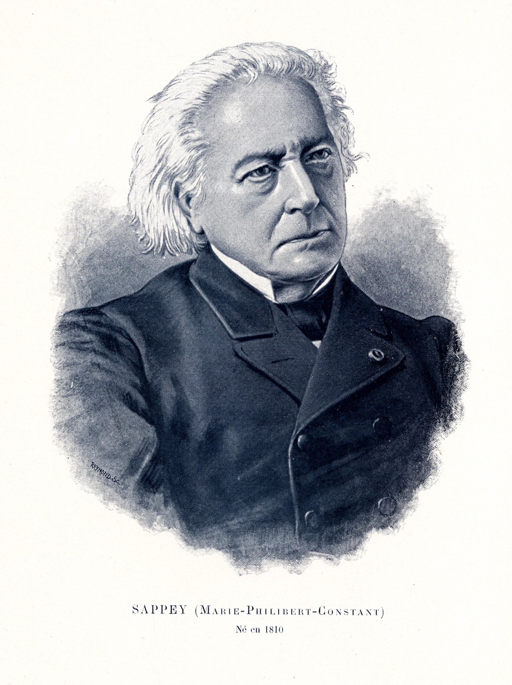 |
| Sappey, Marie Philibert Constant (1894) The author of the image is S. Reymond? (see Corlieu A. Centenaire de la Faculté de Médecine de Paris (1794-1894). Paris, 1894.); original in the BIU Santé Médecine collection, image: CIPN21614 (Licence Ouverte / Open License, color correction) |
Keywords
ligamentum capitis femoris, ligamentum teres, ligament of head of femur, role, significance, anatomy, blood supply, vascularization, stroma, microanatomy, structure
.
NB! Fair practice / use: copied for the purposes of criticism, review, comment, research and private study in accordance with Copyright Laws of the US: 17 U.S.C. §107; Copyright Law of the EU: Dir. 2001/29/EC, art.5/3a,d; Copyright Law of the RU: ГК РФ ст.1274/1.1-2,7
Comments
Post a Comment