Fragments from
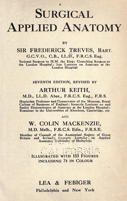
the book Treves F, Keith A, Mackenzie C. Surgical Applied Anatomy, 7th ed. (1917).
The authors discusses the strength and significance of the ligamentum capitis
femoris (LCF) and its changes in hip dislocations and dysplasia.
Quote pp. 542-543
3. THE HIP-JOINT
…
The manner in which
the various movements at the hip are limited may be briefly expressed as follows:
The limit of every natural movement is fixed by the extensibility of the muscles
which surround a joint. That is readily seen at the hip-joint, for when the knee
is extended, and the hamstring muscles thus tightened, flexion at the hip is limited
long before the ligaments become tense. Ligaments only come into play when the muscular
defence of the joint breaks down. Flexion, when the knee is bent, is limited by
the contact of the soft parts of the groin. Extension, by the ilio-psoas, rectus
femoris, and the ilio-femoral or Y -ligament. Abduction, by the adductor mass of
muscles and the pubo-capsular ligament. Adduction of the flexed limb, by the gluteal
musculature, the ligamentum teres and ischio-capsular ligament. Rotation outwards
is resisted by the tensor fasciae femoris, the anterior parts of the glutei medius
and minimus, and the ilio-femoral ligament. The ligamentum teres, which is not a
strong ligament, becomes taut when the thigh is flexed and rotated outwards. It
is ruptured in all cases of complete dislocation. The structures which take the
chief part in maintaining the integrity of the joint, however, are not the ligaments
but the strong muscles which surround and act on the joint. Atmospheric pressure
bears no part, for the fat at the transverse notch is readily drawn into the acetabulum
to make good any space vacated by the femoral head in all normal movements erf the
hip-joint. All joints are provided with yielding pads of fat to obviate alterations
of atmospheric pressure interfering with movements in the joint.
Quote p. 550
Blood is brought
to the head of the femur by vessels in the neck of the bone and in the reflected
parts of the capsule, but only an insignificant supply is carried by the ligamentum
teres (Walmsley). A deficiency in the blood supply to the fragments has been often
put forward to explain failures of union, but there is no real evidence to support
this contention.
Quote pp. 553-554
Congenital dislocation
of the hip-joint is due in most instances to a failure in the development of the
acetabulum. In such cases the acetabulum retains the shallow character seen during
the second month of foetal life. The outgrowth of the acetabular rim fails, especially
in the iliac part. The acetabular cavity becomes filled up by the duplication of
the capsule, which is unduly lax (Fig. 123). The round ligament may be intact or
deficient. The head of the femur becomes flat and the neck short, and the bone slips
backwards on the dorsum ilii when the child learns to walk. The weight of the body
is supported by the muscles and ligaments round the hip-joint, and the gait of the
patient resembles the waddle of a duck. If replaced the head again slips from the
shallow cavity. In time osteophytic outgrowths from the ilium lead to the formation
of a new cavity. The deformity is evidently correlated with the development of the
female sexual organs, for it occurs nearly nine times more frequently in female
than in male children (Fairbanks).
Quote pp. 554-555
General facts. —
In all these dislocations of the hip, (a) the luxation occurs when the limb is in
the position of abduction; (b) the rent in the capsule is always at its posterior
and lower part; (c) the head of the bone always passes at first more or less directly
downwards; (d) the Y-ligament is untorn, while the ligamentum teres is ruptured.
(a) It is maintained that, in all luxations at the hip, the pelvis and femur are in the mutual position of abduction of the latter at the time of the accident. The lower and inner part of the acetabulum is very shallow, and the lower and posterior part of the capsule is very thin. In abduction the head of the bone is brought to the shallow part of the acetabulum; it moves more than half out of that cavity; it is supported only by the thin weak part of the capsule, and its further movement in the direction of abduction is limited only by the pubo-capsular ligament, a somewhat feeble band. In abduction the round ligament is slack, and in abduction with flexion both the Y-ligament and the ischio-capsular ligaments are also relaxed. In the position of abduction, therefore, no great degree of force may be required to thrust the head of the bone through the lower and posterior part of the capsule and displace it downwards.
External links
Treves F, Keith
A, Mackenzie C. Surgical Applied Anatomy. 7th ed. Philadelphia, New York: Lea
and Febinger, 1917. [archive.org]
Authors &
Affiliations
Frederick
Treves (1853–1923) was a British surgeon and anatomist. [wikipedia.org]
 |
| Frederick Treves (1884) Unknown author, original in the wikimedia.org collection (CC0 – Public Domain, no changes). |
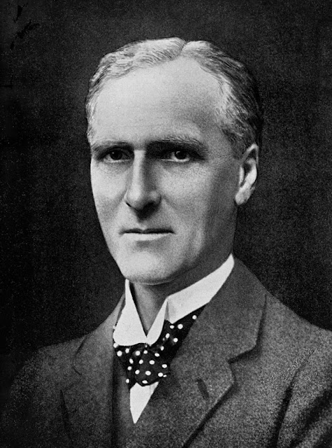
Arthur Keith (1866-1955)
was a British anatomist and anthropologist. [wikipedia.org]
 |
| Arthur Keith Unknown author and date, original in the wikimedia.org collection (CC BY 4.0, no changes). |
Colin Mackenzie (1877-1938) was an Australian anatomist. [wikipedia.org]
Keywords
ligamentum capitis femoris, ligamentum teres, ligament of head of femur, anatomy, role, significance, dislocation, dysplasia
NB! Fair practice / use: copied for the purposes of criticism, review, comment, research and private study in accordance with Copyright Laws of the US: 17 U.S.C. §107; Copyright Law of the EU: Dir. 2001/29/EC, art.5/3a,d; Copyright Law of the RU: ГК РФ ст.1274/1.1-2,7

